



Fusion pull-down plus mass spectrometry (MS) detects a protein–protein interaction only as co-recovery. A fusion tag occupies a matching resin. The cargo is presented because it is fused. Proteins that remain through a written wash travel with that fusion onto the bead, are eluted, digested, and become peptide identifications. The mechanism of detection is this conversion of a wash-stable association into a reporter signal. Calling the signal a binding site, a binding-strength value, or a cellular network collapses that conversion.
The tag can be glutathione S-transferase (GST), His, Strep, biotin, or Flag. The resin is glutathione (GSH), nickel or cobalt, streptavidin-class, or a tag ligand. An antibody against the tag is usually named immunoprecipitation, not pull-down. An empty-fusion arm is part of the mechanism: it says which identifications were already explained by the tag or the resin. For the two linked events named without this detection chain, Principle of Fusion Protein Interaction Analysis Using Pull-Down and MS provides a complementary principle.
Researchers who already have a tagged fusion and want this detection chain can also review the MtoZ Biolabs Pull Down based Protein Analysis Service with Mass Spectrometry for project-specific feasibility and analysis planning.
Tag Occupancy Starts Detection
Detection starts with a tag that can occupy beads in the chosen buffer. Genetic fusion adds the handle. It does not guarantee that the cargo still folds or still presents the intended face. A GST, His, or Flag fusion that is insoluble, or that is a tag stub, still occupies the resin and still presents whatever remains.
The ligand on the bead recognizes the tag, not the prey. Prey enters the chain only if it stays with the cargo. For antibody capture, where the first occupancy is an epitope rather than a fusion tag, Mechanism of Co-Immunoprecipitation in Detecting Protein-Protein Interactions provides a complementary Co-IP detection chain.
The Fusion Complex Is Occupancy, Not Purity
Once the tag is bound, the complex on the bead is the fusion, the resin, and whatever still occupies the cargo or the handle. Partners are not recognized by GSH, nickel, or streptavidin unless they themselves carry that tag. They remain if their off-rate is slower than the incubation and wash in that buffer. Mild detergent and 4 °C slow dissociation. They do not preserve every weak pair, and they do not prevent post-lysis contacts from forming.
Washing is a kinetic filter. It is not elution of “non-specific proteins” that leaves a high-purity complex. Proteins that bind the tag, the linker, or the resin survive the same filter.
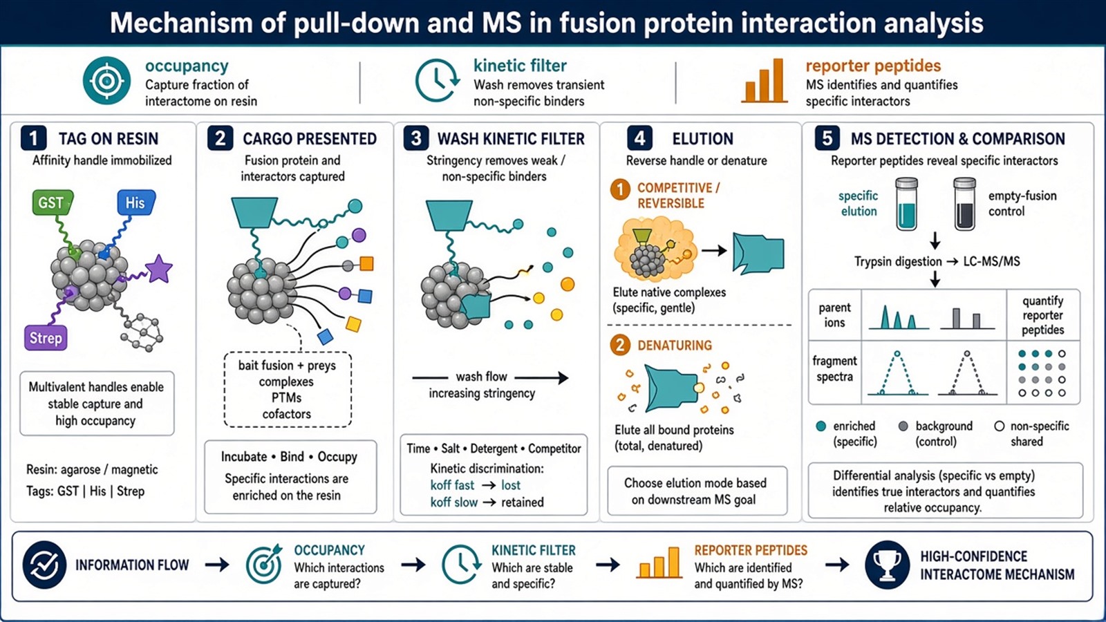
Figure 1. Tag occupancy, cargo presentation, wash as a kinetic filter, elution, then digestion and fragment spectra. The interaction itself is not the measured quantity.
Name which bond will be reversed at elution before the first wash. Competitive ligand and SDS do not release the same mixture.
Elution Reverses One Bond
A competitive ligand, such as free GSH or imidazole, reverses tag–resin occupancy and releases the fusion together with whatever still rides on it. Acid or SDS denatures the assembly and can release more protein, including more handle binders. Both exits produce a mixture. Neither exit sorts cargo binders from tag binders. Fusion and empty-fusion must use the same exit.
MS Is the Reporter, Not a Second Mechanism of Binding
Trypsin converts the recovered proteins into peptides. Electrospray ionization, or another ionization route, puts those peptides into the spectrometer. Liquid chromatography reduces co-elution. Sequence assignment comes from fragment spectra against a database. Parent mass-to-charge values do not identify a protein. Intact-complex molecular weight is not measured. Amino-acid sequence of the whole protein is not read continuously.
Quantification, when a label or matched runs are added, reports relative occupancy after wash. It does not report binding strength, a change in binding site, or the assembly order of a complex. Those sentences need a different mechanism. For how advantages and limits follow from this chain, Advantages and Disadvantages of Pull-Down and MS in Fusion Protein Interaction Analysis provides a complementary decision page.
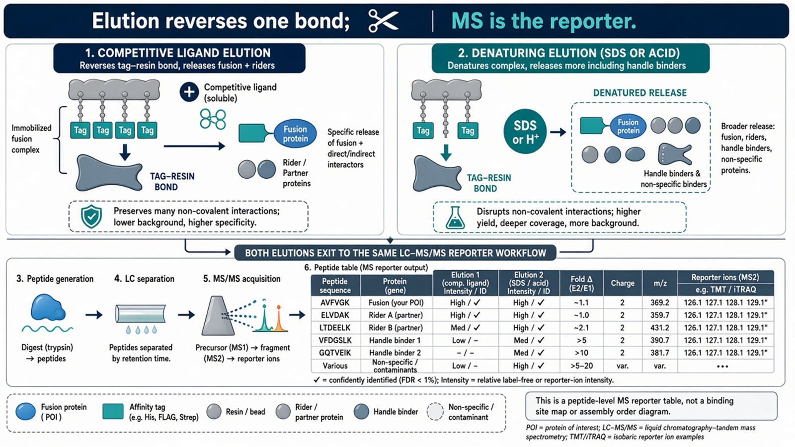
Figure 2. Competitive elution reverses the handle. Denaturing elution breaks more contacts. Empty-fusion uses the same exit. The reporter is a peptide table, not a structural mechanism.
What This Mechanism Does Not Convert
Identification of accessions in the fusion arm, enriched against empty-fusion, is the licensed product. Confirming a nominated pair, or listing unnominated candidates, stays inside that product.
Comparing two conditions can change who remains after wash. That is a change in occupancy. It is not an investigation of interaction mechanisms at the residue level and not a construction of a cellular interaction network. Signal transduction, cell-cycle, and metabolic drawings are later overlays. For GST-specific coupling after this chain is named, GST Pull-Down Assay Combined with Mass Spectrometry for Protein-Protein Interaction Analysis provides a complementary GST–MS page.
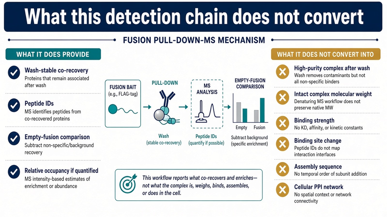
Figure 3. Supported: wash-stable co-recovery reported as peptides. Not converted by this mechanism: binding site, assembly sequence, intact mass of the complex, or a pathway map.
Experimental Limits
The mechanism reduces a mixture to what the tag, the resin, the wash, and the search engine can report. Diagnostic language sits outside ordinary research reporting unless a separately validated assay framework is in place.
Project Planning and Service Scope
A usable mechanism plan names the tag, the bond that elution will reverse, the empty-fusion arm, and the claim that will be allowed, before the first induction. MtoZ Biolabs scopes fusion pull-down with Western or Orbitrap-class LC-MS/MS. Interpretability still follows this chain, not the resin brand.
Once the handle is specified as GST, the wet capture can be scoped as GST pull-down. Researchers who need a GST-bait plan before identification can review the MtoZ Biolabs GST Pull Down Assay Service for project-specific sample evaluation, feasibility assessment, and workflow planning.
Frequently Asked Questions
1. What is the mechanism of fusion pull-down plus MS?
A tag occupies resin, wash-stable proteins ride the fusion, elution releases a mixture, and MS converts that mixture into peptide identifications against empty-fusion.
2. Does washing yield a high-purity complex?
No. Washing is a kinetic filter. Tag and resin binders can remain. Empty-fusion estimates that residue.
3. Can MS after pull-down report binding sites or assembly order?
No. It reports peptide-supported accessions and, if quantified, relative occupancy. Sites and assembly order need a different method.
4. How does this page differ from the principle page?
The principle page names tag occupancy plus peptide identification. This page follows the detection chain: which bond is occupied, which bond is reversed, and what the reporter can say.
5. Is an antibody against the tag a pull-down?
The bead logic is similar. The name and the control are usually IP, with an isotype or empty-tag IP, not a ligand-only resin.
Reference
- Smith D.B., Johnson K.S. (1988). Single-step purification of polypeptides expressed in Escherichia coli as fusions with glutathione S-transferase. Gene, 67, 31-40. https://doi.org/10.1016/0378-1119(88)90005-4
- Gingras A.C., Gstaiger M., Raught B., Aebersold R. (2007). Analysis of protein complexes using mass spectrometry. Nat. Rev. Mol. Cell Biol., 8, 645-654. https://doi.org/10.1038/nrm2208
- Dunham W.H., Mullin M., Gingras A.C. (2012). Affinity-purification coupled to mass spectrometry: basic principles and strategies. Proteomics, 12, 1576-1590. https://doi.org/10.1002/pmic.201100523
- Vikis H.G., Guan K.L. (2004). Glutathione-S-transferase-fusion based assays for studying protein-protein interactions. Methods Mol. Biol., 261, 175-186. https://doi.org/10.1385/1-59259-762-9:175
- Phizicky E.M., Fields S. (1995). Protein-protein interactions: methods for detection and analysis. Microbiol. Rev., 59, 94-123. https://doi.org/10.1128/mr.59.1.94-123.1995
