



RNA immunoprecipitation (RIP) is used to investigate RNAs associated with a selected RNA-binding protein or other RNA-associated protein in cells or tissues. An antibody captures the target protein together with its associated ribonucleoprotein (RNP) complexes, allowing the recovered RNA to be analyzed by RT-qPCR or sequencing. RIP-qPCR is suited to testing specific RNA candidates, whereas RIP-seq enables broader identification of associated RNAs. The choice of assay depends on whether the research objective is targeted validation or transcriptome-wide discovery.
Researchers investigating broader interaction strategies involving proteins, DNA, RNA, and other molecules can explore Protein Interaction Analysis. For studies centered on a known protein and its associated RNAs, the RNA Immunoprecipitation (RIP) Service can support experimental planning and downstream RNA analysis. If you need to identify or validate RNAs associated with a target protein, explore MtoZ Biolabs’ RNA Immunoprecipitation (RIP) Service.
How RIP Investigates RNA Associated with a Known Protein
RIP starts with a protein of interest, commonly an RNA-binding protein or another protein suspected of participating in RNA-associated complexes. Cells or tissues are lysed under conditions designed to preserve relevant molecular associations. An antibody against the target protein is then incubated with the lysate, and the resulting immune complexes are captured using beads or another suitable support. After washing to reduce nonspecific material, the recovered complexes are processed to release and purify RNA for downstream analysis.
Early ribonomics studies established antibody-based approaches for isolating endogenous mRNA populations associated with specific RNA-binding proteins (Tenenbaum et al., 2002). Since then, RIP has been used to investigate candidate RNA–protein associations and to identify broader sets of RNAs associated with a selected protein.
The downstream assay determines the scope of the analysis:
- RIP-qPCR: Tests whether selected RNA transcripts are enriched in the target-protein immunoprecipitate relative to appropriate controls. It is particularly useful when candidate RNAs are already known.
- RIP-seq: Uses sequencing to identify a broader set of RNAs recovered with the target protein, supporting candidate discovery and transcriptome-wide analysis.
- RIP-Chip: Combines immunoprecipitation with microarray-based RNA profiling to examine associated RNA populations (Keene et al., 2006).
Some RIP workflows include crosslinking to stabilize molecular associations before lysis, whereas others preserve complexes under non-crosslinking conditions. Crosslinking can help retain interactions that are otherwise difficult to preserve, while also affecting recovery and background. The choice should reflect the target protein, sample type, and research objective.
RIP can recover RNAs that directly contact the target protein, as well as RNAs associated indirectly through other proteins or larger RNP complexes. This distinction is important when interpreting the results and deciding which follow-up experiments are needed.
When RIP Is an Appropriate Method
RIP is particularly suitable when the protein of interest is known and the research question concerns which RNAs associate with that protein. Common applications include testing whether a candidate transcript is enriched with a specific RNA-binding protein, comparing RNA associations across treatment conditions, and identifying associated RNA populations on a broader scale.
Successful experiments depend on the presence of the target protein in the selected biological material, the availability of a suitable antibody, and the recovery of RNA of sufficient quality for downstream analysis. Antibody performance is especially important: an antibody that detects a protein by Western blot does not necessarily capture it efficiently or specifically during immunoprecipitation.
RIP is less suitable when the starting point is a known RNA and the aim is to discover unknown proteins. That reverse question is addressed by RNA Pull-Down Assay. RIP is also not a substitute for mapping chromatin associated with an endogenous RNA; that occupancy question requires a chromatin-isolation design such as ChIRP. RIP is not the preferred route for mapping a precise protein contact site at nucleotide resolution. Crosslink-based methods designed for contact mapping may be more appropriate when site resolution is essential.
Biological context also matters. The target protein and RNA should be present in the selected sample and, where relevant, in compatible cellular compartments. For example, an RNA predominantly localized in the cytoplasm may be poorly represented in an experiment focused on a nuclear protein under conditions that do not preserve their association. Expression patterns, subcellular localization, treatment conditions, and developmental state can all influence the RNAs recovered.
Choosing Controls and Ensuring Sample Quality
Appropriate controls help distinguish target-associated RNA enrichment from nonspecific recovery and background signals. Their selection should reflect the target protein, immunoprecipitation strategy, and likely sources of background.
- Input: An aliquot collected before immunoprecipitation provides a reference for the RNA present in the starting material and supports interpretation of enrichment.
- IgG control: A matched nonspecific immunoglobulin control helps assess background associated with antibody and bead interactions.
- Target-specific controls: Depending on the experimental system, knockout or knockdown samples, untagged controls, or other suitable negative controls can provide additional evidence of specificity.
Antibody specificity is a central determinant of interpretability. A western blot should show whether the target is present, but immunoprecipitation performance should be assessed directly because epitope accessibility differs between assays. Heavy nonspecific capture can obscure true enrichment even when the target protein is recovered.
RNA integrity must also be maintained from sample collection through RNA purification. Degradation can reduce transcript detection, distort abundance profiles, and compromise sequencing library preparation. Cells and tissues should be collected and stored under appropriate conditions, with handling procedures designed to limit RNase contamination.
Sample requirements depend on the biological material, target protein abundance, antibody performance, and downstream assay. These factors should be considered together when determining the amount and condition of material needed for a study.
Biological replicates are important when assessing reproducibility or comparing experimental conditions. Treatment and control groups should be processed consistently, and batch effects should be minimized so that biological differences are not confounded with differences in extraction, immunoprecipitation, or library preparation.
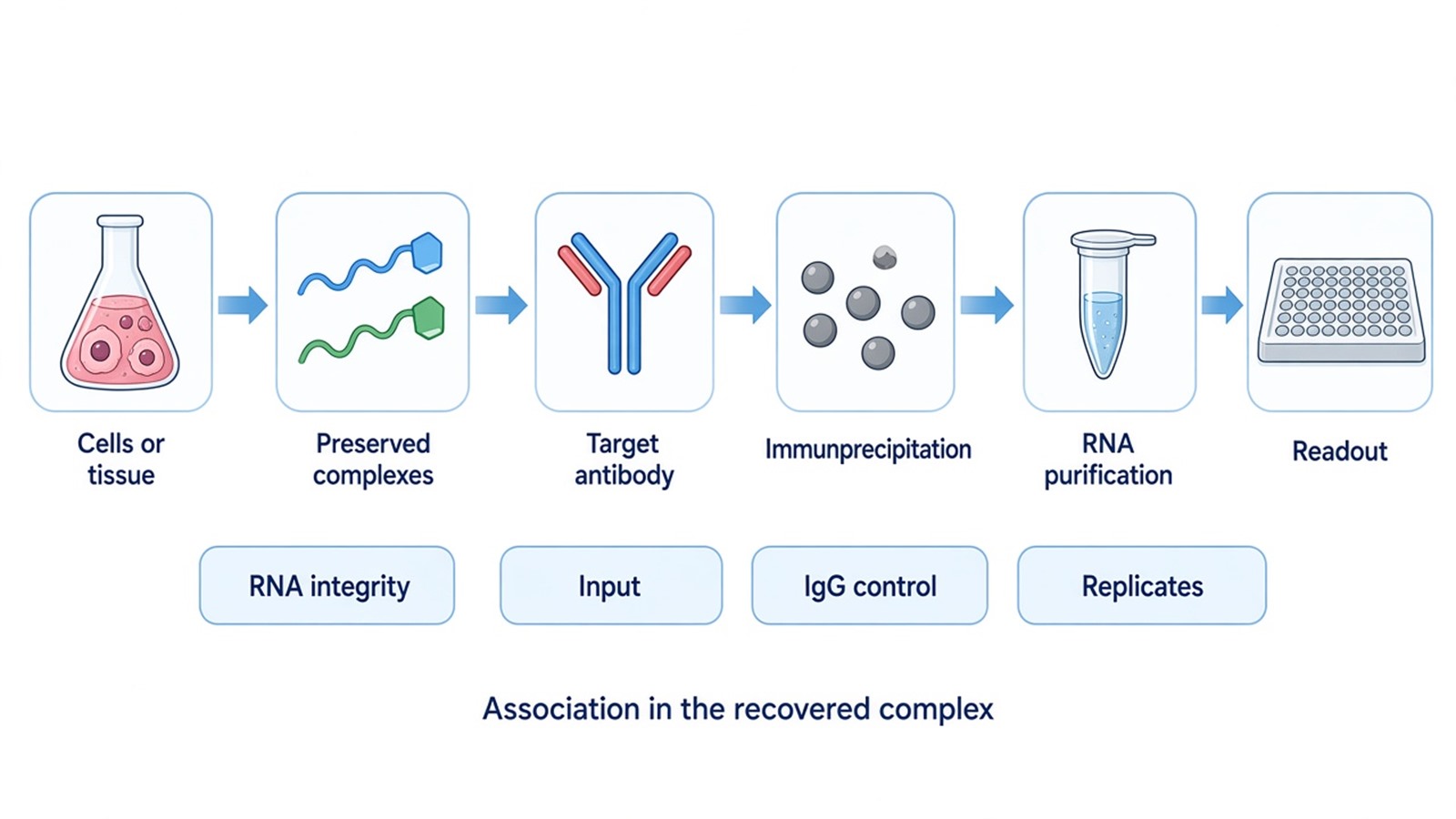
Figure 1. RIP interpretation depends on antibody specificity, RNA integrity, matched controls, and a readout aligned with the research question.
Interpreting RIP-qPCR and RIP-seq Results
RIP-qPCR is commonly interpreted through enrichment relative to input and negative control. A candidate RNA that is reproducibly enriched in the specific immunoprecipitation has evidence of association with the recovered protein complex. The magnitude should be considered together with RNA abundance, amplification quality, control behavior, and replicate consistency. A large numerical enrichment from a weak or variable signal is not automatically stronger evidence.
RIP-seq generates a broader list or profile of RNAs associated with the target protein. Analysis may include read quality assessment, alignment, transcript-level or region-level enrichment, comparison with input and IgG controls, and functional annotation of candidate RNA sets. Statistical significance does not eliminate experimental background, so candidates should be reviewed for abundance, reproducibility, localization, and biological plausibility.
RIP-seq signal should not be interpreted as a precise binding site unless the experimental and analytical method was specifically designed to support such resolution. A recovered region may reflect transcript abundance, fragmentation, complex organization, or analysis choices. Likewise, enrichment of an RNA does not demonstrate that the protein changes its stability, splicing, localization, or translation.
Independent validation can include RIP-qPCR on selected sequencing candidates, reciprocal RNA-centered capture, altered protein expression, or functional assays. The follow-up should test the next claim rather than merely reproduce the same enrichment format. For example, if the claim concerns direct recognition of an RNA element, purified-component binding or a site-sensitive method is more informative than another broad RIP.
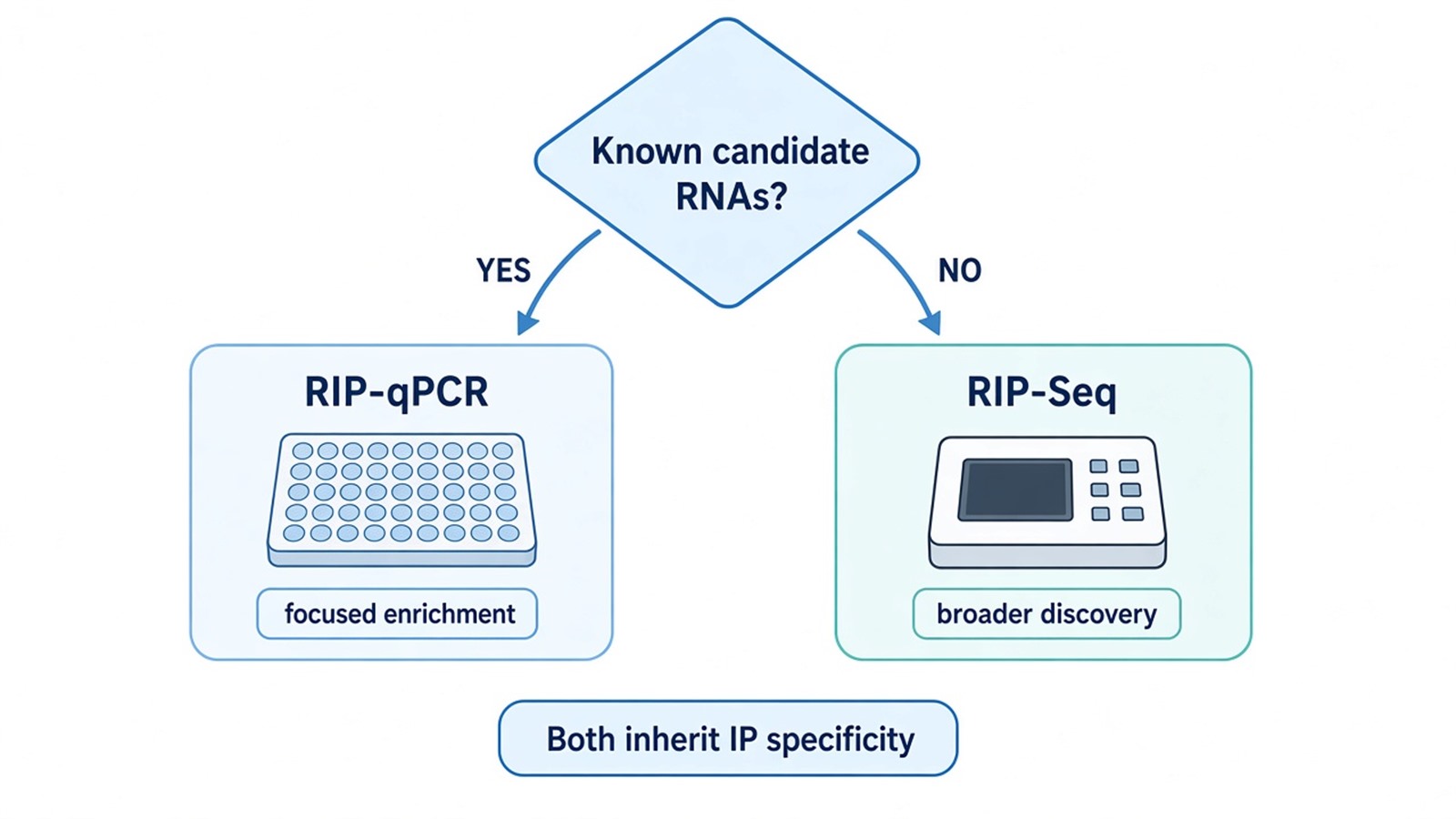
Figure 2. RIP-qPCR suits focused candidate testing, whereas RIP-Seq supports broader discovery; both inherit the specificity and reproducibility of the immunoprecipitation.
Frequently Asked Questions
1. Can an antibody that works for western blotting be used for RIP?
Not necessarily. Western blotting often recognizes denatured protein, whereas RIP requires effective capture under the chosen lysis and immunoprecipitation conditions. Antibody specificity and target recovery should be evaluated in an immunoprecipitation context. If the target cannot be enriched cleanly, downstream RNA data will be difficult to interpret.
2. Is it better to submit frozen cells, tissue, or purified RNA?
For a full RIP workflow, cells or tissue preserve the starting protein-RNA complexes. Purified RNA alone cannot support immunoprecipitation of the protein complex. RNA purified after a completed immunoprecipitation may be suitable for downstream qPCR or sequencing if quality and quantity are adequate. Sample type, preservation, and transport should be reviewed before collection.
3. What can be done when the IgG control has substantial RNA signal?
First determine whether the signal comes from abundant input RNA, nonspecific antibody binding, beads, or incomplete washing. A more specific antibody, an alternative bead system, optimized lysis and wash conditions, or an additional biological specificity control may help. The remedy should target the identified source of background rather than relying only on a higher numerical cutoff.
4. Should RIP-qPCR or RIP-seq be selected for a first experiment?
RIP-qPCR is usually more focused when a small set of candidate RNAs already exists and assayable primers can be designed. RIP-seq is appropriate when the RNA repertoire is unknown and broader discovery is justified. A pilot confirmation of target protein recovery and RNA quality can reduce uncertainty before a larger sequencing design.
5. Does RIP enrichment prove that the protein regulates the RNA?
No. Enrichment supports association with the recovered complex. A regulatory claim requires additional evidence, such as perturbing the protein and measuring a relevant RNA processing, stability, localization, translation, or phenotypic outcome. Direct contact and functional consequence remain separate questions.
Conclusion
RIP provides a practical approach for investigating RNA populations associated with a known protein. Reliable results depend on suitable antibody performance, preservation of RNA quality, appropriate controls, and a downstream assay matched to the research objective. RIP-qPCR is useful for targeted testing, while RIP-seq supports broader discovery of protein-associated RNAs.
If you are planning an RIP study, contact MtoZ Biolabs with your target protein, sample type, research objective, and any candidate RNAs or preliminary results. This information can help guide a discussion of the appropriate RIP strategy and downstream analysis.
Reference
- J.D. Keene, J.M. Komisarow, M.B. Friedersdorf (2006). RIP-Chip: the isolation and identification of mRNAs, microRNAs and protein components of ribonucleoprotein complexes from cell extracts. Nature Protocols, 1, 302-307. https://doi.org/10.1038/nprot.2006.47
- S.A. Tenenbaum, P.J. Lager, C.C. Carson, J.D. Keene (2002). Ribonomics: identifying mRNA subsets in mRNP complexes using antibodies to RNA-binding proteins and genomic arrays. Methods, 26, 191-198. https://doi.org/10.1016/S1046-2023(02)00022-1
