



A washed human cell pellet is sent for proteomic identification when the laboratory needs a named protein inventory from that isolate. Typical material is a cell-line or primary-cell pellet after culture or isolation. Bottom-up liquid chromatography-tandem mass spectrometry (LC-MS/MS) extracts proteins, digests them to peptides, and matches fragment spectra to a human sequence database under a declared false discovery rate (FDR). The identification list reports protein groups supported by accepted peptides.
Harvest, residual medium, lysis completeness, and the search database decide which names appear. The list does not, by itself, report copy number, pathway activity, or a group fold change. Host cell protein (HCP) impurity testing, single-cell proteomics, and endogenous peptidomics use different starting material and different claims.
What a Human Cell Protein Identification List Reports
An identification list is a protein-group table. Peptides are scored from fragment spectra, filtered at a peptide-level FDR, and rolled up to proteins that share peptide evidence (Nesvizhskii and Aebersold 2005; Elias and Gygi 2007). A row usually means that at least one accepted peptide was assigned to that group under the declared search settings. It does not mean that every isoform, splice form, or proteoform of the gene was distinguished.
Unique peptides support one protein group. Shared peptides can pull homologs into the same row or split one gene product across several rows. Keratin, residual trypsin, and bovine serum albumin from fetal bovine serum (FBS) should be searched in a contaminant database so they are labeled as carryover rather than as unexpected human hits (Mellacheruvu et al. 2013).
A human cell identification list reports accepted protein groups, not an amount, a localization, or a function.
Researchers who need how cell extracts move from lysis to database search can refer to Cell Proteomics: Principles, Workflow, and Applications.
How Harvest and Lysis Decide Which Proteins Enter the Digest
The usual order for human cell protein identification is fixed: a defined pellet, a PBS wash, lysis, digestion, LC-MS/MS, a human database search, and an identification list. Each step removes or retains a different protein set. Medium and FBS left on the pellet become high-intensity bovine proteins. Culture trypsin left after detachment becomes a dominant enzyme hit. Incomplete lysis leaves membrane and nuclear proteins in the debris.
Adherent lines are commonly detached with culture trypsin or a scraper. Residual culture trypsin should be minimized before the pellet is frozen, because that enzyme is later indistinguishable from digestion trypsin except by experimental record. Suspension cells are pelleted and washed. Primary isolates and mixed immune populations need the same wash discipline, plus a record of cell type and isolation method, because a mixed pellet produces a mixed inventory.
Lysis chemistry decides solubility. Detergent or urea-based buffers recover more membrane proteins than a mild native buffer. A whole-cell lysate reports proteins that entered that extract. Nuclear, membrane, or secretome claims require a declared fractionation or collection route. Snap-freezing and dry-ice shipment protect the pellet that was actually harvested.
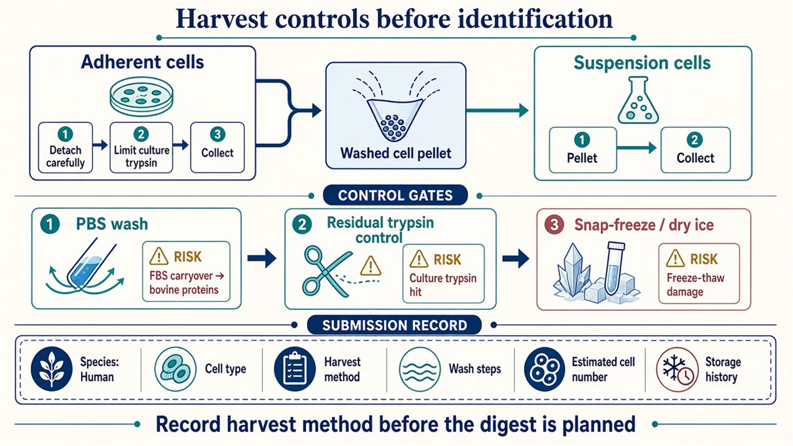
Figure 1. Wash, residual trypsin, freeze-thaw, and harvest method should be recorded before a human cell pellet is treated as identification-ready.
Collection, storage, and shipping details for cell pellets are summarized in Preparing Cell Samples for Proteomics Analysis: Collection, Storage, and Handling.
Factors That Change Identification Depth Before Search
Identification depth is the number of protein groups that pass the declared FDR after a given digest and acquisition. Cell input, contamination, lysis, and search settings move that number before any bioinformatics ranking is applied.
|
Factor |
What it changes in the list |
What to record with the pellet |
|---|---|---|
|
Cell input and protein load |
Detectable peptide count and recovery of lower-abundance groups |
Estimated cell number or protein amount |
|
FBS or medium carryover |
Bovine albumin and other serum proteins occupy signal |
Wash steps and medium composition |
|
Residual culture trypsin |
A high-intensity enzyme hit and extra cleavage products |
Detachment method and residual-trypsin control |
|
Lysis buffer and debris removal |
Recovery of membrane, nuclear, or cytoskeletal proteins |
Buffer class and whether the lysate was clarified |
|
Database and isoforms |
Which human accessions and shared-peptide groups appear |
Swiss-Prot versus a larger isoform set |
|
Contaminant database |
Whether keratin, trypsin, and BSA are labeled as carryover |
Contaminant library used in the search |
|
Enzyme rule and FDR |
Peptide acceptance and protein-group size |
Missed cleavages, specificity, peptide and protein FDR |
Routine cellular proteomics projects are often planned around approximately 5 × 10^6 cells per sample as a working minimum and approximately 1 × 10^7 cells when a deeper inventory is requested. Low-input pellets can be evaluated, but the expected list is then limited by material rather than by search software. Those ranges are planning values. They are not a promised protein count.
How to Judge Whether the Project Is Identification-Only
An identification-only project fits when the scientific question is “which proteins are supported in this human cell extract.” A quantitative project fits when predefined groups must be compared in one matrix. A named-protein project fits when the list is already closed and a scheduled assay is required.
|
Scientific need |
Design that fits |
Do not treat the ID list as |
|---|---|---|
|
Open inventory from a human pellet |
Discovery LC-MS/MS identification |
Proof that unlisted proteins are absent |
|
Same-protein comparison across groups |
Label-free, DIA, or TMT quantitative design |
A substitute fold-change table |
|
Closed protein names |
Targeted PRM or MRM in a new digest |
Discovery coverage of unscheduled proteins |
|
Biologic impurity monitoring |
An HCP-specific method |
A human cell-line inventory |
|
Endogenous peptides without routine trypsin |
Peptidomics |
Shotgun protein inference |
|
One-cell or ultra-low input |
A dedicated low-input workflow |
A standard whole-pellet ID list |
HCP analysis asks which production-host proteins remain in a purified biologic. Human cell protein identification asks which proteins are supported in a research cell pellet. The two projects share LC-MS/MS hardware and do not share sample definition, database intent, or reporting units.
If the pellet is already collected and the question is still an open inventory, keep the project on discovery identification. If groups, treatments, or time points must be compared, lock a quantitative design before the first digest is queued. Researchers who already have human cell pellets and a stated identification or comparison goal can also review the MtoZ Biolabs Cellular Proteomics Service for project-specific harvest and search planning.
Sample Planning for Cell-Line and Primary Pellets
Cell-line pellets are usually homogeneous enough that one harvest record can represent the culture. Primary cells, stem-cell derivatives, and sorted immune populations need a tighter record of isolation, viability, and remaining feeder or serum components. Mixed starting material produces a mixed identification list even when the search database is correctly human.
A usable submission record names species as human, cell type, culture or isolation background, treatment if any, harvest method, wash steps, estimated cell number, and storage history. Cell pellets are the preferred submission format when extraction still needs to be matched to the project. Frozen lysates and extracted protein can be accepted when those steps are already locked and documented. Snap-freeze in liquid nitrogen or store at −80 °C, ship on dry ice, and keep freeze-thaw cycles to one or two.
Adherent and suspension harvests should stay consistent across a cohort if later quantification is possible. Changing scraper versus culture trypsin, or changing wash count, between “control” and “treated” pellets creates list differences that later software cannot assign to biology.
Interpretation Limits of an Identification List
Presence on the list means accepted peptide evidence under the stated FDR, database, and digestion rule. Absence from the list means the pipeline did not report a usable match. Low-abundance proteins, shared-peptide collapse, aggressive filters, and incomplete lysis can all hide a protein that is biologically present (Nesvizhskii and Aebersold 2005).
The list does not support an amount or concentration claim. Intensity-based ranks inside one run can help plan a follow-up assay. They are not calibrated copy numbers. Group fold change requires a quantitative design with matched extraction and a declared contrast. Pathway enrichment on an identification-only table describes annotation of detected names. It does not demonstrate pathway activity.
Subcellular localization is outside a whole-cell identification list unless a fractionation step was declared. Personalized-medicine or diagnostic cutoffs sit outside ordinary research reporting. Disease-mechanism language should stay limited to proteins that the design actually measured.
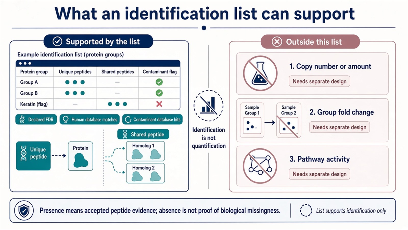
Figure 2. An identification list can support protein groups, human-database matches, and contaminant labels; amount, fold change, and pathway activity need another design.
Frequently Asked Questions
1. Is host cell protein analysis the same project as human cell protein identification?
No. HCP work monitors residual production-host proteins in a purified biologic. Human cell protein identification reports protein groups from a research cell pellet searched against a human database and a contaminant set.
2. How many cells are typically planned for a routine identification project?
Routine cellular proteomics is often planned at about 5 × 10^6 cells as a working minimum and about 1 × 10^7 cells when a deeper list is requested. Low-input pellets need a separate evaluation. Cell number still does not guarantee a fixed protein count.
3. Should protein be extracted before the pellet is shipped?
No as a default. A washed, snap-frozen pellet lets extraction be matched to the identification plan. Pre-extracted protein is usable when the lysis buffer and protein amount are already documented.
4. Does an identification list report relative abundance between groups?
No. The list reports accepted protein groups. Relative comparison requires a quantitative acquisition and analysis plan. Targeted confirmation is a separate digest when names are already closed.
5. Why do bovine serum proteins appear in a human cell search?
FBS and residual medium remain on poorly washed pellets and produce strong bovine peptide matches. A contaminant database labels those hits. Additional PBS washes before freezing reduce that carryover.
MtoZ Biolabs can provide proteomic identification of human cell proteins from cell-line or primary pellets, including harvest-to-search planning, human-database identification, and review of residual medium or trypsin effects when project samples and study design are available.
If further evaluation of cell input, lysis route, or whether the project should stay identification-only is needed, project details can be submitted for a feasibility review.
Reference
- A.I. Nesvizhskii, R. Aebersold (2005). Interpretation of shotgun proteomic data: the protein inference problem. Mol. Cell. Proteomics, 4, 1419-1440. https://doi.org/10.1074/mcp.R500012-MCP200
- J.E. Elias, S.P. Gygi (2007). Target-decoy search strategy for increased confidence in large-scale protein identifications by mass spectrometry. Nat. Methods, 4, 207-214. https://doi.org/10.1038/nmeth1019
- D. Mellacheruvu, Z. Wright, A.L. Couzens, J.P. Lambert, N.A. St-Denis, T. Li (2013). The CRAPome: a contaminant repository for affinity purification-mass spectrometry data. Nat. Methods, 10, 730-736. https://doi.org/10.1038/nmeth.2557
- J.R. Wisniewski, A. Zougman, N. Nagaraj, M. Mann (2009). Universal sample preparation method for proteome analysis. Nat. Methods, 6, 359-362. https://doi.org/10.1038/nmeth.1322
- Y. Zhang, B.R. Fonslow, B. Shan, M.C. Baek, J.R. Yates 3rd (2013). Protein analysis by shotgun/bottom-up proteomics. Chem. Rev., 113, 2343-2394. https://doi.org/10.1021/cr3003533
