



The principle of SILAC-based Co-IP-MS is a mass pair on a Co-IP eluate, not a more specific antibody. Cells incorporate light or heavy lysine and arginine while they grow. Co-immunoprecipitation (Co-IP) still captures a bait and the proteins that survive the written wash. Mass spectrometry (MS) then sees the same peptide at two masses and reports a heavy-to-light (H/L) ratio. That ratio ranks recovery under a written contrast. It does not report an absolute copy number, a binding constant, or a direct contact. Stable isotope labeling by amino acids in cell culture (SILAC) does not change the three Co-IP events. It adds a second channel so that proteins that entered after mixing can be separated from proteins that were already bait-dependent.
The useful first decision is when the two lysates are mixed. Mix after two separate IPs and the ratio includes handling differences between tubes. Mix before one IP and bead or antibody contaminants tend toward ratio 1, while partners that were already on the bait keep the biological imbalance. Confusing those two mix points is how a SILAC-Co-IP-MS table is over-read.
Researchers who already have a proliferating cell system, an IP-grade antibody or tag, and a written light–heavy contrast can also review the MtoZ Biolabs Co-IP-MS Protein Interaction Analysis Service for project-specific feasibility and analysis planning.
What SILAC Adds to Co-IP-MS
SILAC is metabolic labeling. Light medium supplies natural lysine and arginine. Heavy medium typically supplies Lys8 and Arg10, or a medium pair such as Lys4 and Arg6 when a third state is used. After several doublings, commonly five to eight, newly synthesized protein carries the label. Incorporation must be checked before the Co-IP is queued. Residual light peptide in the heavy channel compresses every later ratio toward 1. Arginine-to-proline conversion can split the heavy pair. Dialyzed serum and medium free of unlabeled Lys and Arg are part of the principle, not optional extras.
Co-IP remains antibody or tag capture plus Protein A/G collection. For those three events without a label, Principle of Co-Immunoprecipitation in Protein Interaction Analysis provides a complementary mechanism discussion. SILAC does not raise antibody specificity. A weak antibody still returns bead binders. They simply appear as a ratio.
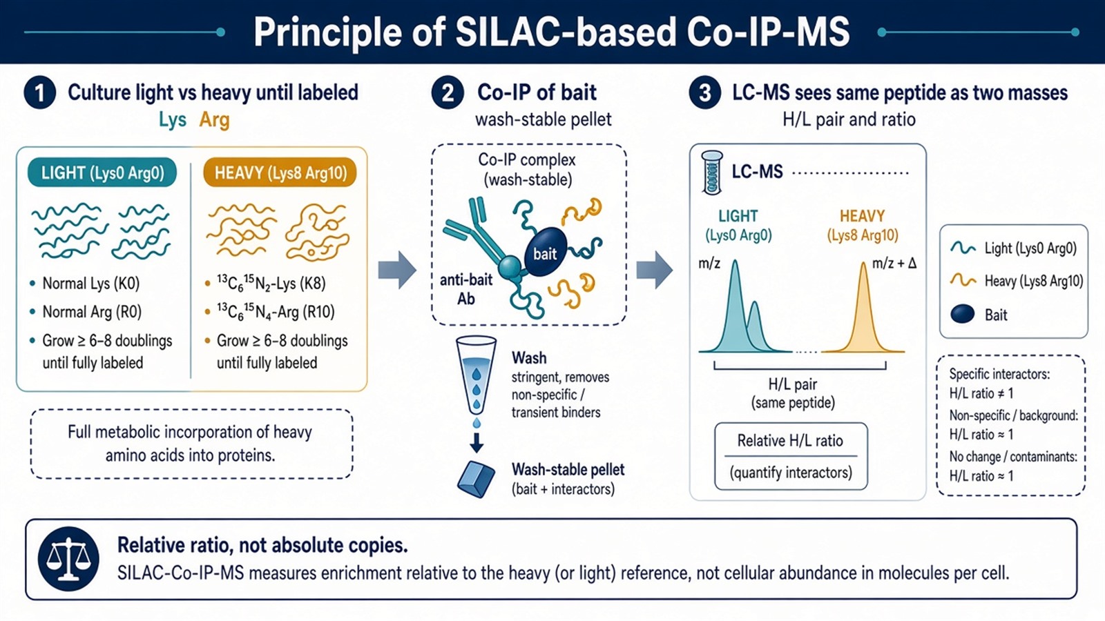
Figure 1. The label is in the protein before lysis. Co-IP collects a wash-stable pellet. MS reads the same peptide as two masses. The principle is a relative H/L ratio, not an absolute quantity.
Write the contrast and the mix point before the first harvest. A ratio cannot invent a control that was not labeled.
Principles of the Coupled Workflow
The original four steps are the same chemistry read as a principle, not as a protocol list.
1. SILAC labeling
Two (or three) populations grow until the proteome is labeled. The two states must name one contrast: bait versus empty-tag or isotype, or the same bait with and without a stimulus. Two contrasts in one unlabeled list cannot be recovered later. Non-dividing primary cells and most tissues cannot be SILAC-labeled in culture. They need a different quantitative route.
2. Cell lysis and Co-IP
Lysis must keep the intended complex, as in unlabeled Co-IP. The principle choice is the mix point. In a mix-after-IP design, each state is captured separately and the eluates are combined before digestion. Tube-to-tube IP differences enter the ratio. In a mix-before-IP design, lysates are combined and one antibody capture is performed. Proteins that bind beads or Fc after mixing occupy both channels and approach H/L ≈ 1. Proteins that were already on the bait in only one state keep an unbalanced ratio. That discrimination is the I-DIRT-class use of the same SILAC principle. Label-swap, forward and reverse, tests whether an outlier is chemistry or a channel artifact.
3. Protein digestion and mass spectrometry analysis
The mixed eluate is digested, usually with trypsin, which cleaves after Lys and Arg and therefore places the SILAC mass difference on most peptides. LC-MS/MS distinguishes the pair by mass-to-charge. The instrument does not measure interaction. It measures paired peptide intensity. Incomplete labeling, missed cleavage, and arginine-to-proline conversion distort that pair.
4. Data analysis
Proteins are ranked by H/L, not by identification alone. Bait should show the expected channel occupancy. Contaminants in a mix-before-IP design cluster near 1. Outliers are candidates for bait-dependent or condition-dependent recovery. They are still wash-stable associations. For how to read that table as specificity versus occupancy, and when TMT or label-free quantification is the better label, Quantitative Analysis of Protein-Protein Interactions Using SILAC and MS provides a complementary ratio discussion. For the four-step analysis path, SILAC-Based Protein-Protein Interaction Analysis Using Mass Spectrometry provides a complementary workflow.
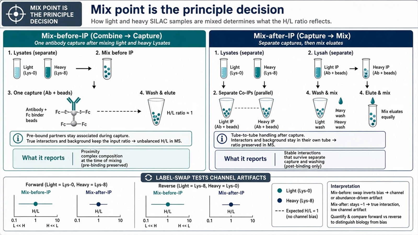
Figure 2. Mix lysates, then one Co-IP: post-mix contaminants approach H/L 1. Mix two finished IPs: handling differences remain in the pair. Label-swap tests channel artifacts.
Detailed Principles
Relative, not absolute, quantification
SILAC compares two labeled states of the same peptide. It is relative quantification. Absolute copy number needs a spike-in standard that was never part of this principle. “Minimizing random error” is true only for steps after mixing. Everything before the mix point, including labeling completeness and unmatched lysis, still enters the ratio.
Specificity is not a SILAC property
Co-IP enriches a pellet. MS identifies what was in that pellet. The label only pairs the two states. High sensitivity of the spectrometer does not convert a ratio-1 bead binder into a partner, and it does not convert an outlier into a direct contact.
Dynamic occupancy is a contrast, not a built-in advantage
A treated-versus-control SILAC-Co-IP can report that a prey’s recovery changed. That is occupancy under the stated stimulus. It is not a movie of the complex, and it is not a regulatory mechanism. A single time point remains a single ratio.
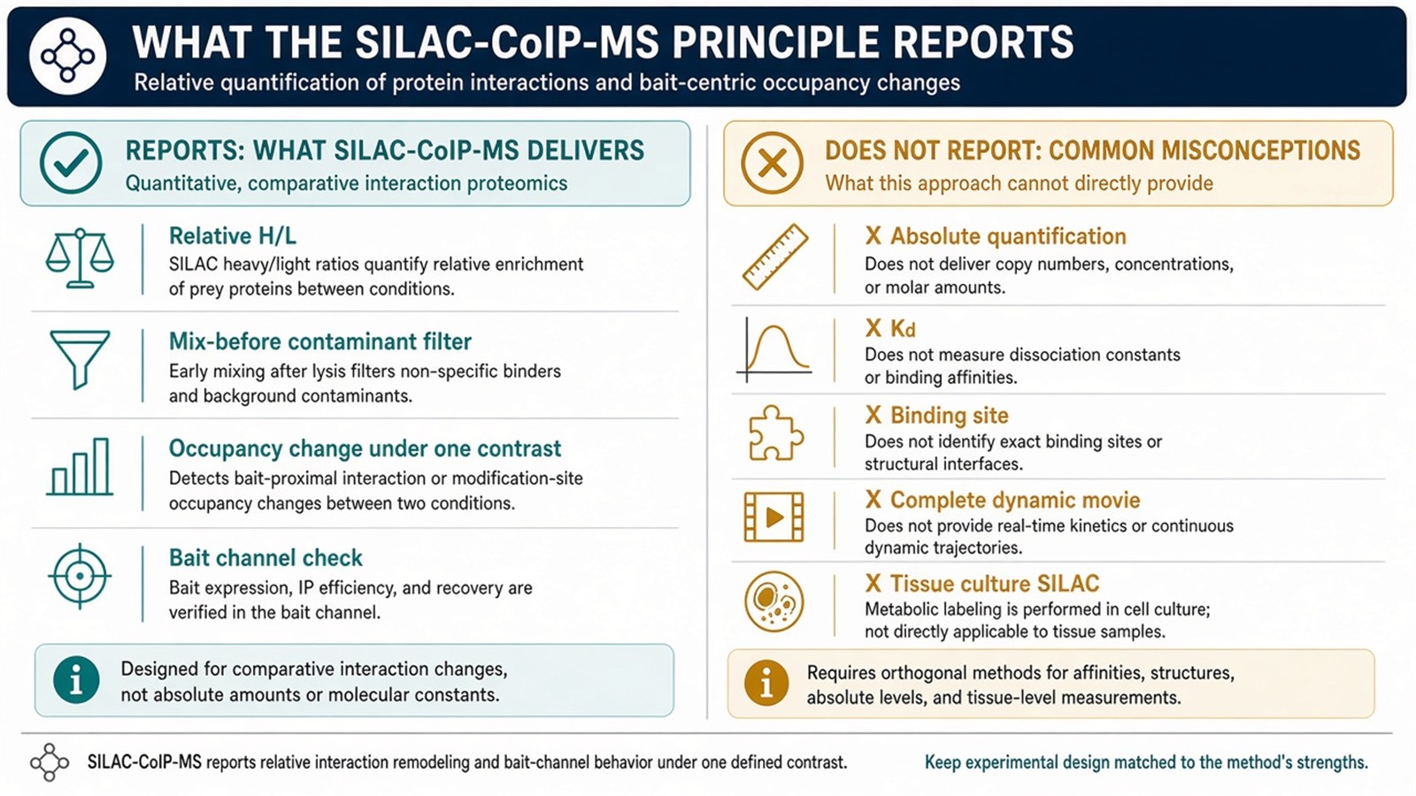
Figure 3. What the principle supports: paired intensity, mix-before-IP contaminant filtering, occupancy change under one contrast. What it does not: absolute abundance, a binding site, or a reconstructed interactome.
Experimental Limits
The principle requires dividing cells, a complete label, a written mix point, and a Co-IP that recovered the bait. Three replicates do not repair a 70% heavy channel. Diagnostic language sits outside ordinary research reporting unless a separately validated assay framework is in place.
Project Planning and Service Scope
A usable SILAC-Co-IP-MS plan names the contrast, the Lys/Arg pair, the incorporation check, the mix point, the Co-IP control, and how outliers will be ranked, before the first medium change. MtoZ Biolabs scopes labeled Co-IP-MS on Orbitrap-class LC-MS/MS when those items are written. Interpretability still follows the ratio and the mix point, not the instrument name.
Once the principle is specified, the project can be run as bait-versus-control or as treated-versus-control Co-IP-MS. Researchers who need a quantitative interactome plan after that decision can review the MtoZ Biolabs Protein-Protein Interaction Analysis Service for project-specific sample evaluation, feasibility assessment, and workflow planning.
Frequently Asked Questions
1. What is the principle of SILAC-based Co-IP-MS?
Metabolic light and heavy proteins are captured by Co-IP and read as paired peptides. The H/L ratio ranks recovery under a written contrast. The interaction itself is not the measured quantity.
2. Does SILAC provide absolute quantification of interactors?
No. It provides a relative ratio between two labeled states. Absolute copies need a designed standard.
3. Why does mix-before-IP push contaminants toward ratio 1?
Proteins that bind beads or Fc after the lysates are combined occupy both channels. Proteins already on the bait in one state do not.
4. Can tissues be used in this principle?
Not by culture SILAC. Tissue or non-dividing samples need another quantitative label or a spike-in design.
5. How does this page differ from other SILAC-MS articles?
This page states why the mass pair and the mix point work. How to read H/L as specificity versus occupancy, and the four-step analysis path, are separate jobs.
Reference
- Ong S.E., Blagoev B., Kratchmarova I., et al. (2002). Stable isotope labeling by amino acids in cell culture, SILAC, as a simple and accurate approach to expression proteomics. Mol. Cell. Proteomics, 1, 376-386. https://doi.org/10.1074/mcp.M200025-MCP200
- Blagoev B., Kratchmarova I., Ong S.E., et al. (2003). A proteomics strategy to elucidate functional protein-protein interactions applied to EGF signaling. Nat. Biotechnol., 21, 315-318. https://doi.org/10.1038/nbt790
- Tackett A.J., DeGrasse J.A., Sekedat M.D., et al. (2005). I-DIRT, a general method for distinguishing between specific and nonspecific protein interactions. J. Proteome Res., 4, 1752-1756. https://doi.org/10.1021/pr050168c
- Trinkle-Mulcahy L., Boulon S., Lam Y.W., et al. (2008). Identifying specific protein interaction partners using quantitative mass spectrometry and bead proteomes. J. Cell Biol., 183, 223-239. https://doi.org/10.1083/jcb.200805092
- Vermeulen M., Hubner N.C., Mann M. (2008). High confidence determination of specific protein-protein interactions using quantitative mass spectrometry. Curr. Opin. Biotechnol., 19, 331-337. https://doi.org/10.1016/j.copbio.2008.06.001
