



Mass spectrometry TMT technology is the way a liquid chromatography-tandem mass spectrometry (LC-MS/MS) instrument reads isobaric tandem mass tags after labeled peptides have been mixed (Thompson et al. 2003). At the MS1 level, channels that share a kit remain one precursor. Fragmentation releases reporter ions whose intensities become relative peptide, then protein, values. TMT technology in mass spectrometry names that readout. It does not name a method that separates differently tagged peptides in a survey scan, and it does not name a test that distinguishes single-amino-acid variants by the tag itself. Sequence ions identify the peptide. Reporter ions quantify the channels. The valid claim is relative change of the same protein inside one multiplex set, not an absolute concentration and not a diagnostic marker.
Researchers who already have defined samples, comparison groups, or a specific project objective can also review the MtoZ Biolabs TMT Quantitative Proteomics Analysis Service for project-specific feasibility and analysis planning.
What the Mass Spectrometer Must Record
TMT technology starts after digestion and amine labeling. The mixed digest is separated by liquid chromatography and ionized. The mass spectrometer has two jobs that must stay linked: assign the peptide and extract channel reporters from the same precursor family. If those two records come from unlinked software trails, the ratio is no longer a TMT measurement.
MS1 cannot split TMT channels. The balancer keeps tags isobaric, so a stronger MS1 peak is not a higher channel. Treating the survey scan as the quantitative TMT signal confuses unlabeled peak-area LFQ with isobaric tagging.
Researchers who need to determine whether that tagging strategy matches a study can refer to TMT-Based Proteomics for more detailed guidance.
Mass spectrometry TMT technology reads reporter ions from a shared precursor, not channel-specific MS1 peaks.
MS2 Reporter Ions and Isolation Interference
In a typical data-dependent acquisition (DDA) TMT method, a precursor is isolated and fragmented. The product-ion spectrum contains peptide fragments used for identification and low-mass reporter ions used for quantification. Isolation is never infinitely narrow. Co-isolated peptides can contribute their own reporters and pull ratios toward one, an effect known as ratio compression (Ting et al. 2011).
Isolation width, chromatographic peak density, and sample complexity set how often that mixing happens. High resolving power can separate close TMTpro reporter pairs that differ by a few millidaltons. It does not remove reporters that came from a different peptide inside the same isolation window. A complete channel matrix can still hide compressed biology. Known isotopic impurities in each reagent lot are corrected in processing. That correction is part of reading the tag. It is not a substitute for a clean isolation window.
In MtoZ isobaric-labeling offerings, TMT methods are typically acquired as DDA so that reporters can be read from the isolated precursor. Data-independent acquisition (DIA) is not a TMT technology synonym. Service configuration is not an academic rule that other laboratories must copy.
When MS3 Changes the Reporter Population
Synchronous precursor selection MS3 (SPS-MS3) adds a second fragmentation: selected peptide fragments from MS2 are isolated and fragmented again, and reporters are read from that cleaner ion population (McAlister et al. 2014). The technology point is a purer reporter spectrum, not a new labeling chemistry and not a higher plex number.
MS3 reduces compression when co-isolation is the main error. It does not repair incomplete labeling, a confounded channel map, or a sample that arrived after mixing. It does not create an amount. Instruments that cannot run SPS-MS3 can still run MS2 TMT. The claim stays relative either way. A lower-complexity digest, or a contrast that can tolerate some compression, may stay on MS2 to keep cycle time for identification. Scan choice is a trade between reporter purity and how many precursors are sampled in the same chromatographic peak.
When compression and closed-plex limits change the experimental plan, researchers can refer to Advantages and Disadvantages of TMT-Based Quantitative Proteomics Analysis for a more detailed strategy-selection framework.
Scan record |
What it contributes |
What it cannot do |
|---|---|---|
MS1 precursor |
One isobaric peak for the mixed peptide |
Split TMT channels or report a channel ratio |
MS2 product ions |
Peptide identity plus reporter intensities |
Exclude all co-isolated reporters |
SPS-MS3 reporters |
Reporters from a selected fragment population |
Recover unlabeled or late-arriving samples |
High resolving power |
Separate close reporter masses |
Remove isolation mixing |
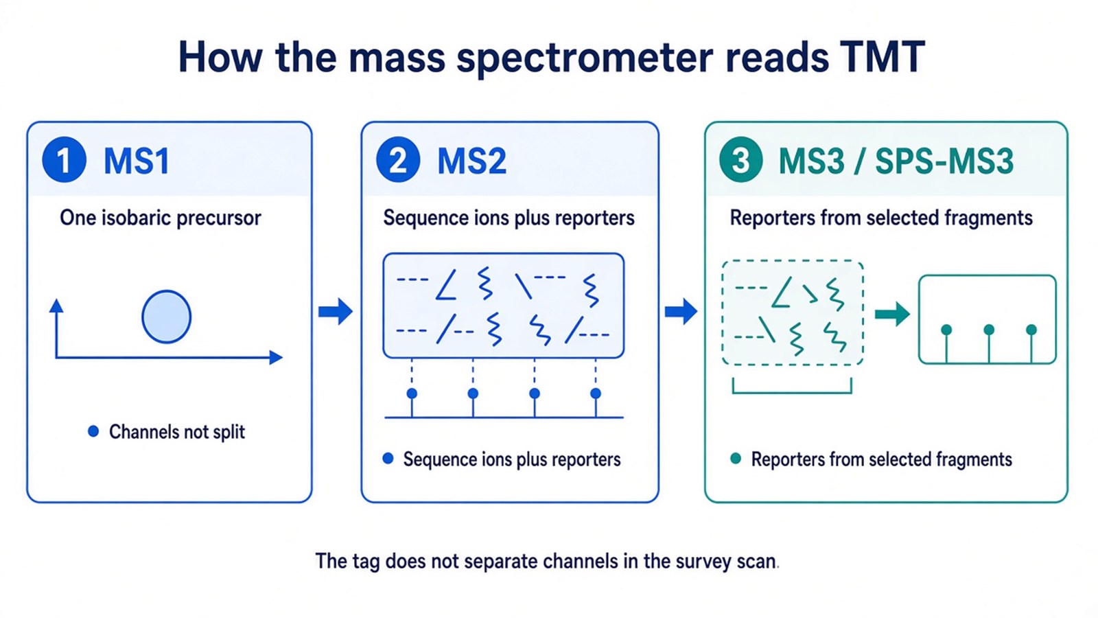
Figure 1. TMT technology in the mass spectrometer is a shared MS1 precursor plus reporter readout at MS2 or MS3; the tag does not split channels in the survey scan.
What Mass Spectrometry TMT Technology Is Not
TMT technology is not a general peptide-separation method. Liquid chromatography separates the digest. The tag does not pull one channel to a different retention time. TMT technology is not a single-residue sequencing tool. Amino-acid differences appear, if at all, as different peptides or site-specific mass shifts, not as a property of the reporter.
TMT technology is not unlabeled discovery with a faster scanner. Independent LFQ files still compare MS1 chromatograms. TMT files compare reporters from a pool. Mixing those descriptions produces tables that are read as the wrong experiment.
Oncology, neuropathology, or drug-response titles do not change the readout. Those studies still need a closed plex and a relative claim. Early diagnosis and personalized treatment are not functions of the scan type.
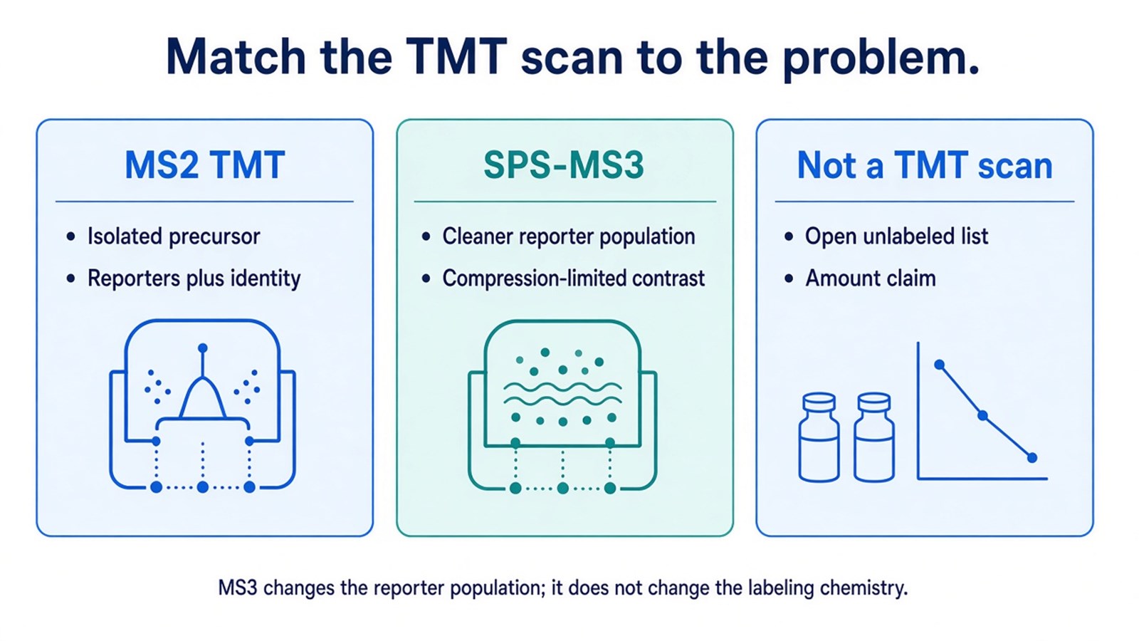
Figure 2. MS2 TMT reads reporters from the isolated precursor; SPS-MS3 is chosen when cleaner reporters are required; an open list or an amount claim is not a TMT scan choice.
A TMT mass-spectrometry record supports relative channel ratios from a chosen scan level, not a proteome-wide concentration or a clinical map.
Frequently Asked Questions
1. Does TMT technology separate labeled samples in the mass spectrometer?
Not at MS1. Channels are isobaric until fragmentation. Separation of samples happens chemically before mixing. The mass spectrometer then reads reporters from the shared precursor.
2. Is MS2 TMT a different technology from MS3 TMT?
Both use the same labels. MS3 changes where reporters are recorded. It is a scan option for compression, not a second reagent class. The protein table should state whether ratios came from MS2 or MS3.
3. Can TMT technology quantify single amino-acid variants because the tag is so specific?
No. The tag reports channel intensity of the labeled peptide that was identified. Variant or modification claims need the corresponding peptide as the analyte.
4. Is TMT technology the same as TMT-based proteomics?
TMT-based proteomics is the labeling study. Mass spectrometry TMT technology is the MS1-isobaric, reporter-ion readout that makes that study measurable. The names are often used together. They still do not mean an unlabeled peak-area experiment.
Once the plex map, intended scan level, and expected protein-level readout are defined, the project can be evaluated as a TMT mass-spectrometry study. Researchers can review the MtoZ Biolabs TMT Quantitative Proteomics Analysis Service for project-specific sample evaluation, feasibility assessment, and workflow planning.
