



An intracellular protein question usually arrives with a washed cell pellet and a spatial claim still unnamed. The laboratory may need a deeper inventory of proteins released from inside the cell, or it may need to assign proteins to nucleus, cytosol, mitochondria, endoplasmic reticulum, or membrane-enriched fractions. Bottom-up liquid chromatography-tandem mass spectrometry (LC-MS/MS) can support either job only when the extract or fraction matches the claim. A whole-cell lysate reports proteins that entered that extract. It does not, by itself, prove organelle residence.
Harvest severity, detergent choice, fractionation purity, and marker checks decide what “intracellular” means in the final table. Host cell protein impurity testing, surface-proteome biotinylation, and endogenous peptidomics use different starting material and different reporting units.
What Intracellular Protein Proteomics Is Asked to Deliver
Intracellular protein proteomics by mass spectrometry asks which proteins are supported in an intracellular extract or in a declared compartment-enriched fraction under a false discovery rate (FDR). The analytical object is still a peptide-to-protein match after digestion. The scientific object is the spatial scope of that extract.
A whole-cell intracellular inventory is useful when the question is coverage of proteins recovered after cell disruption, including many organelle proteins that become soluble once membranes are opened. A subcellular design is required when the question names a compartment or asks whether a protein redistributed after treatment. Those two endpoints share LC-MS/MS hardware and do not share sample preparation.
Protein groups on an identification list report accepted peptide evidence (Nesvizhskii and Aebersold 2005). Unique peptides support one group. Shared peptides can merge homologs. Contaminant databases should label keratin, residual trypsin, and serum carryover so they are not read as unexpected intracellular hits.
Researchers seeking a broader understanding of cellular protein identification, quantification, and proteomic profiling can explore MtoZ Biolabs' Cellular Proteomics Service
Whole-Cell Extract Versus Subcellular Fractionation
A whole-cell route gently or strongly lyses the pellet, clarifies debris when required, digests the extract, and searches a species-matched database. Detergent or urea-based lysis recovers more membrane and nuclear proteins than a mild native buffer. The list is an intracellular inventory of that extract. It remains silent on which organelle a protein occupied before lysis.
A subcellular route separates compartments before digestion. Differential centrifugation pellets nuclei at low speed, then mitochondria-enriched material at medium speed, then microsomal and membrane-rich material at higher speed. Density gradients refine fractions that share similar sedimentation behavior. Immunoaffinity or magnetic enrichment targets organelles when a surface marker is available (Itzhak et al. 2016). Each fraction is extracted, digested, and analyzed as its own LC-MS/MS sample or as a quantitative series across fractions.
Marker proteins decide whether a fraction is usable. Nuclear, mitochondrial, cytosolic, and endoplasmic reticulum markers should enrich in the intended fraction and deplete elsewhere. Cross-contamination does not disappear after database search. It becomes a localization error that software cannot retract.
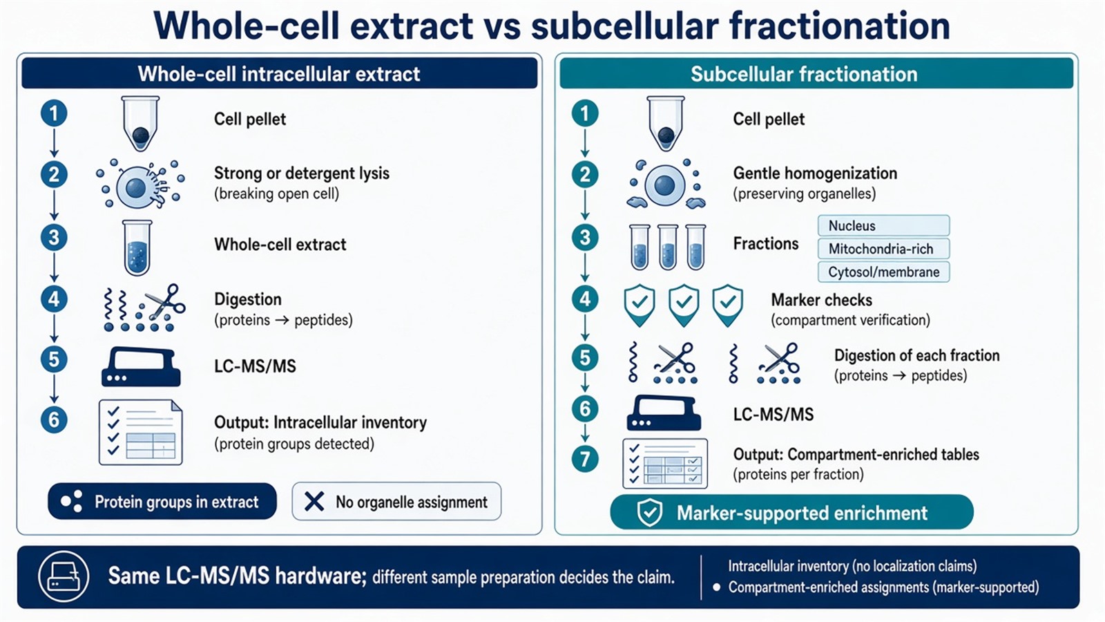
Figure 1. Whole-cell extracts support an intracellular inventory; compartment claims require fractionation and marker checks before LC-MS/MS.
Practical fractionation steps and purity limits are summarized in How to Perform Subcellular Fractionation for Proteomics Analysis?.
Factors That Control Intracellular Coverage and Localization Strength
Coverage is the number of protein groups recovered from an intracellular extract. Localization strength is how confidently a protein can be assigned to a compartment after fractionation. The two metrics move for different reasons.
|
Factor |
Effect on whole-cell inventory |
Effect on compartment assignment |
|---|---|---|
|
Lysis buffer strength |
Detergent or urea recovers more membrane and nuclear proteins |
Harsh lysis before fractionation can rupture organelles |
|
Clarification of debris |
Removes insoluble material from the digest |
Over-clearing can discard intended organelle pellets |
|
Fractionation method |
Not used |
Differential, gradient, or affinity steps set enrichment |
|
Marker validation |
Optional for inventory claims |
Required before localization language is used |
|
Cross-contamination |
Dilutes low-abundance proteins |
Misplaces proteins across compartments |
|
Cell input |
Limits depth of the intracellular list |
Limits how many fractions can be digested |
|
Database and FDR |
Controls accepted protein groups |
Controls names used in localization scoring |
Organelle rupture during homogenization mixes mitochondrial matrix proteins into cytosol-like supernatants. Incomplete nuclear pelleting leaves chromatin-associated proteins in later fractions. Membrane sheets co-sediment with mitochondria when medium-speed spins are too aggressive. Those failures look like biology if marker profiles are ignored.
Quantitative strategies such as label-free, data-independent acquisition (DIA), tandem mass tags (TMT), or stable isotope labeling by amino acids in cell culture (SILAC) can compare the same protein across fractions or conditions (Itzhak et al. 2016). Quantification does not repair a failed fractionation. It reports ratios on whatever material entered the digest.
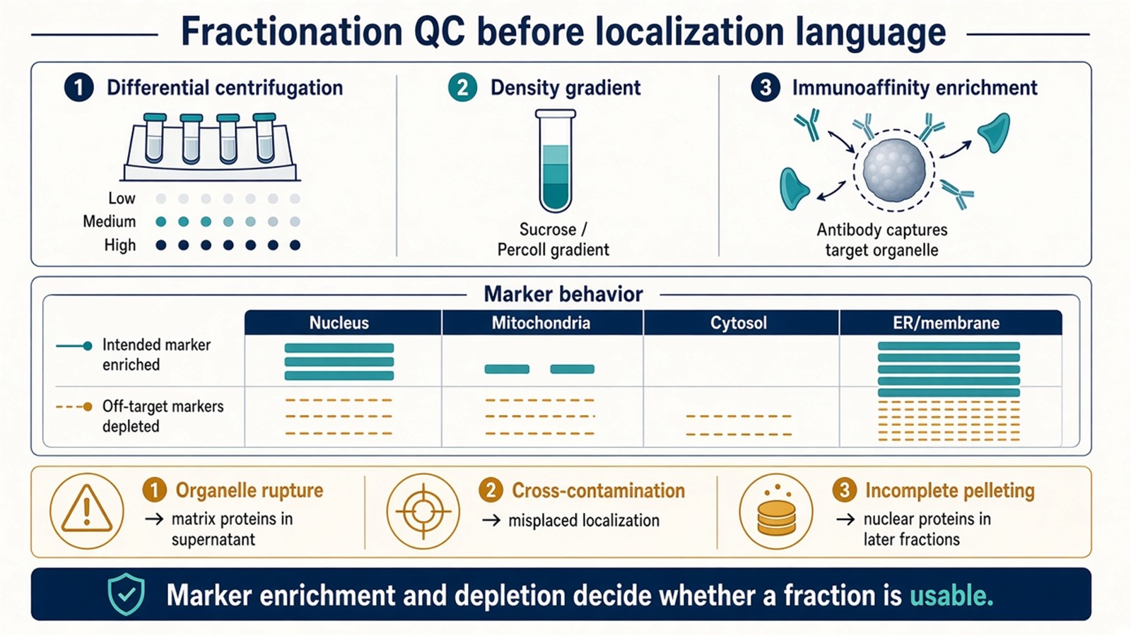
Figure 2. Localization strength depends on fractionation method and marker behavior, not on search depth alone.
How to Judge Which Intracellular Design Fits
The useful test is whether the scientific sentence names a compartment.
|
Scientific need |
Design that fits |
Do not report from this design |
|---|---|---|
|
Deeper inventory of proteins from a disrupted cell |
Whole-cell intracellular LC-MS/MS |
Organelle of origin |
|
Composition of one target organelle |
Fraction enrichment plus marker QC |
Proof of exclusive localization without depletion data |
|
Protein redistribution after treatment |
Matched fractions across conditions, usually quantitative |
Whole-cell fold change as translocation |
|
Closed protein names in one compartment |
Targeted PRM or MRM after enrichment |
Open discovery of unscheduled proteins |
|
Surface-exposed proteins only |
Surface labeling or membrane-selective prep |
Intracellular matrix inventory |
|
Production-host impurity in a biologic |
Host cell protein method |
Research intracellular map |
If the pellet is already collected and the question is still “which proteins are inside this extract,” keep the project on whole-cell intracellular identification or quantification. If the question names nucleus, mitochondria, or translocation, lock fractionation and markers before the first digest is queued. Researchers who already have cultured cells or tissues and a stated compartment goal can also review the MtoZ Biolabs Protein Subcellular Localization Service for fractionation and localization planning.
For how mass spectrometry is paired with organelle enrichment and quantitative readout, researchers can refer to Mass Spectrometry in Subcellular Proteomics: Organelle Fractionation, Localization, and Quantitative Analysis.
Sample Planning for Intracellular and Fractionation Projects
Cultured cells remain the most common starting material for intracellular work because harvest and homogenization can be matched across a cohort. Primary cells and tissues need a tighter record of dissociation, extracellular matrix, and blood or serum carryover. FFPE and heavily fixed material need a separate feasibility review before fractionation is promised.
A usable submission record names species, cell or tissue type, treatment, whether the endpoint is whole-cell inventory or named compartments, lysis or homogenization preference, and storage history. For subcellular localization projects, cell input is often planned at about 1 × 10^7 cells or more so several fractions can be digested. Tissue projects commonly start near 50 mg when fractionation is required. Those values are planning ranges. They are not a promised protein count per organelle.
Protease inhibitors belong in fractionation buffers when endogenous proteases would otherwise digest the intended proteome before the controlled trypsin step. SDS, high glycerol, and extreme salt complicate later cleanup and should be declared if already present. Snap-freeze pellets or tissues, ship on dry ice, and keep freeze-thaw cycles to one or two.
Adherent and suspension harvests should stay consistent across groups if later quantitative comparison is planned. Changing scraper versus culture trypsin, or changing homogenization force, between control and treated arms creates fraction shifts that later software cannot assign to biology.
Interpretation Limits of Intracellular Mass Spectrometry Tables
Presence in a whole-cell intracellular list means accepted peptide evidence in that extract. Absence means the pipeline did not report a usable match. Low-abundance proteins, shared-peptide collapse, and incomplete lysis can hide proteins that remain biologically present.
Presence in one organelle fraction means enrichment under the declared fractionation and marker rules. It does not prove exclusive residence unless depletion in other fractions is shown. Dual-localized proteins and trafficking intermediates can appear in more than one fraction for biological reasons as well as for contamination.
Pathway enrichment on an intracellular identification list annotates detected names. It does not demonstrate pathway activity inside a named organelle. Copy number and absolute amount require matched standards. Diagnostic or personalized-medicine cutoffs sit outside ordinary research reporting.
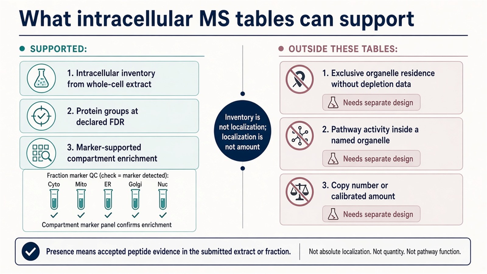
Figure 3. Whole-cell lists support intracellular inventory; compartment language needs marker-checked fractions, and amount or pathway activity needs another design.
Frequently Asked Questions
1. Can a deep whole-cell proteome replace subcellular fractionation?
No. Deeper coverage increases the intracellular inventory. It does not assign proteins to nucleus, mitochondria, or membrane compartments after the cell has been fully lysed.
2. Which fractionation method should be chosen first?
Differential centrifugation fits coarse enrichment of nuclei and mitochondria-rich pellets. Density gradients refine fractions with similar sedimentation. Immunoaffinity enrichment fits organelles with a usable surface marker. Marker QC decides whether the chosen method is enough.
3. How should fraction purity be checked?
Marker proteins should enrich in the intended fraction and deplete elsewhere. Western blot or MS-based marker scoring can document that pattern before localization language is used.
4. Does quantification prove translocation?
Only when matched fractions are compared under a declared design. A whole-cell fold change does not show that a protein moved between compartments.
5. Is intracellular proteomics the same as host cell protein analysis?
No. Intracellular research maps proteins from a cell extract or organelle fraction. Host cell protein work monitors residual production-host proteins in a purified biologic.
MtoZ Biolabs can provide intracellular protein proteomics by LC-MS/MS for whole-cell extracts and for fractionation-based compartment studies, including harvest-to-fraction planning, marker-oriented QC review, and guidance on whether the endpoint should stay inventory-only when project samples and study design are available.
If further evaluation of lysis route, organelle targets, or whether a quantitative localization design is required is needed, project details can be submitted for a feasibility review.
Reference
- A.I. Nesvizhskii, R. Aebersold (2005). Interpretation of shotgun proteomic data: the protein inference problem. Mol. Cell. Proteomics, 4, 1419-1440. https://doi.org/10.1074/mcp.R500012-MCP200
- D.N. Itzhak, S. Tyanova, J. Cox, G.H.H. Borner (2016). Global, quantitative and dynamic mapping of protein subcellular localization. Elife, 5, e16950. https://doi.org/10.7554/eLife.16950
- Y. Zhang, B.R. Fonslow, B. Shan, M.C. Baek, J.R. Yates 3rd (2013). Protein analysis by shotgun/bottom-up proteomics. Chem. Rev., 113, 2343-2394. https://doi.org/10.1021/cr3003533
- J.R. Wisniewski, A. Zougman, N. Nagaraj, M. Mann (2009). Universal sample preparation method for proteome analysis. Nat. Methods, 6, 359-362. https://doi.org/10.1038/nmeth.1322
- T.P.J. Dunkley, R. Watson, J.L. Griffin, P. Dupree, K.S. Lilley (2004). Localization of organelle proteins by isotope tagging (LOPIT). Mol. Cell. Proteomics, 3, 1128-1134. https://doi.org/10.1074/mcp.T400009-MCP200
