



When two proteins bind, which regions become protected, more exposed, or structurally altered? Hydrogen-deuterium exchange mass spectrometry (HDX-MS) addresses this question by comparing deuterium uptake in the unbound protein and protein complex under matched conditions. The resulting peptide-level map helps identify regions affected by binding, prioritize candidate interaction interfaces, and reveal conformational changes beyond the contact surface.
Researchers studying a defined protein pair can use the MtoZ Biolabs Hydrogen Deuterium Exchange Mass Spectrometry Service to discuss construct quality, complex occupancy, and whether a binding-state comparison is feasible. Residue-pair distance restraints from covalent capture are a different evidence type and are covered in Cross-Linking Mass Spectrometry (XL-MS) for Protein Interaction Mapping.
How Does HDX-MS Reveal Binding-Associated Protein Changes?
In HDX-MS, a protein is incubated in deuterated buffer, allowing backbone amide hydrogens to exchange with deuterium. After a defined labeling period, the exchange reaction is quenched, the protein is digested, and peptide deuterium uptake is measured by mass spectrometry. Structured regions with stable hydrogen bonding or limited solvent accessibility generally exchange more slowly than flexible, solvent-exposed regions (Zhang and Smith, 1993). Protein binding can alter these patterns by shielding a surface, stabilizing a local structure, or inducing conformational changes at sites distant from the binding interface (Konermann et al., 2011).
HDX-MS results are interpreted through comparisons between defined states. An unbound protein or an alternative-state control provides a reference for determining which regions change upon complex formation. Multiple labeling times help characterize exchange kinetics, while overlapping peptides can improve regional localization. The resulting spatial resolution is generally at the peptide level rather than the individual-residue level.
HDX-MS is useful for studying proteins that are difficult to crystallize, including some membrane proteins and intrinsically disordered systems, provided that the proteins remain sufficiently stable and the relevant states can be maintained under exchange-compatible conditions.
HDX-MS does not directly measure binding affinity. Deuterium uptake reflects the combined effects of solvent accessibility, hydrogen bonding, protein dynamics, and exchange kinetics. Independent evidence of complex formation, such as a biophysical binding measurement under relevant conditions, helps establish the context for interpreting uptake differences. For a broader overview of complementary evidence types, see Protein-Protein Interaction Analysis: From Partner Discovery to Interaction Validation.
Designing HDX-MS Comparisons for Protein-Protein Binding
Unbound vs Bound States
The core comparison is between the unbound protein and the protein-protein complex. Both states should use matched construct boundaries, buffer composition, temperature, pH, labeling times, and digestion procedures. If the nominally bound sample contains a substantial fraction of free protein, the observed protection may be diluted, making a binding-associated change appear weaker than it actually is. Evidence that the complex forms under the planned experimental conditions should therefore be reviewed before labeling. A previously established interaction can support study design, but complex occupancy under the actual HDX-MS conditions also matters.
The study should specify which protein partner will be mapped. Labeling one protein in the presence and absence of its binding partner identifies regions affected on that protein. Mapping both partners can provide a more complete view of the interface, but requires adequate peptide coverage and reproducible measurements for each protein. Increasing the concentration of one binding partner may increase complex occupancy, depending on the affinity and stoichiometry. However, aggregation or nonspecific association can complicate interpretation, so protein stability should be assessed under the selected conditions.
Experimental Comparability
Differences in pH, temperature, deuterium content, quench timing, or protease activity can produce apparent uptake changes that arise from technical variation rather than binding. Replicate measurements help establish whether peptide-level differences are reproducible. Multiple labeling times can also distinguish persistent protection from changes that emerge only during particular phases of the exchange process.
Before starting a study, confirm that the purified proteins have sufficient purity and quantity for the planned labeling times, replicate measurements, and peptide coverage. Buffer compatibility must be assessed for both complex formation and deuterium exchange. These factors directly affect whether differences between the unbound and bound states can be interpreted reliably.
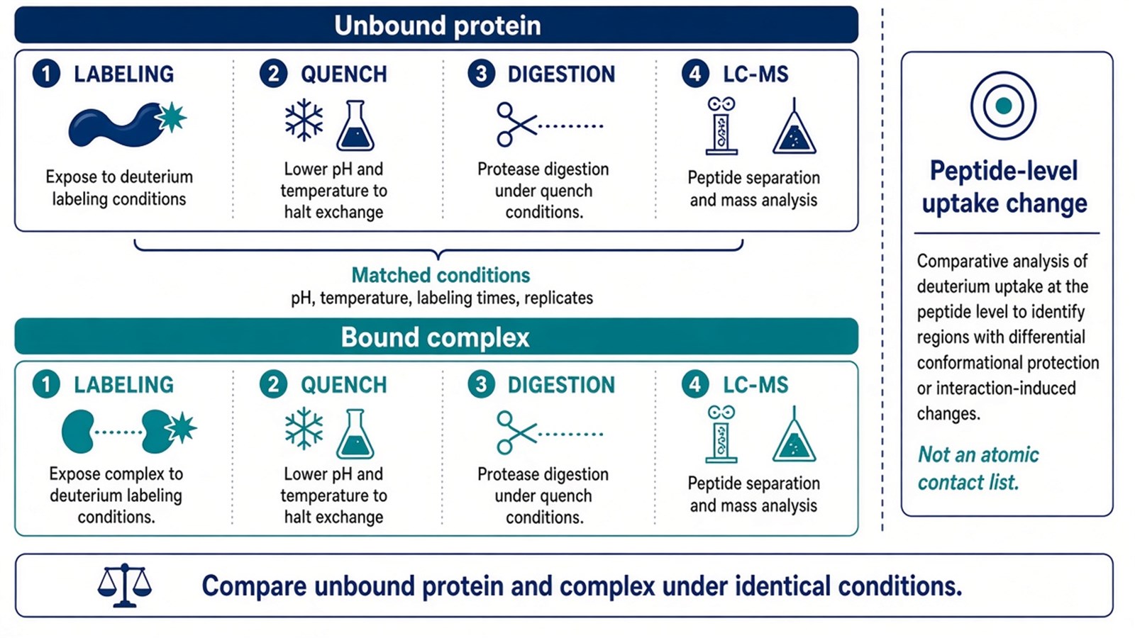
Figure 1. A defensible HDX-MS binding study compares unmatched protein and complex under identical labeling, quench, digestion, and acquisition conditions.
Reading Deuterium Uptake Changes Across Protein Regions
Increased Uptake
Increased deuterium uptake after binding can indicate increased solvent accessibility, greater local flexibility, or destabilization of hydrogen-bonded structure. In a protein-protein system, these changes may reflect an allosteric opening, disruption of an intramolecular contact, or a change in oligomeric state. An increase in uptake is not necessarily inconsistent with binding. The interpretation depends on the affected region, peptide coverage, labeling-time profile, and reproducibility across measurements.
Decreased Uptake
Decreased uptake, commonly called protection, can indicate surface shielding, stabilization of hydrogen bonds, or reduced protein dynamics. When protected peptides cluster on a plausible contact surface, they support a candidate interface. Protection across distant regions may instead reflect a broader conformational response. Protection that develops mainly at later labeling times can indicate slower structural stabilization rather than a permanently buried surface.
Interpretation should remain at the peptide level. Overlapping peptides can narrow the region associated with an uptake change, but do not automatically identify the contacting residue. Missing peptide coverage also limits interpretation: a region without measurable uptake data cannot be classified as unaffected. Contaminants, incomplete digestion, and back-exchange should be evaluated when they could affect the reliability of uptake measurements.
Interpreting HDX-MS Changes at Candidate Interfaces
Candidate Interface Patterns
A candidate interface is more plausible when protected peptides form a spatially coherent region on a potential contact surface and independent evidence supports binding between the same protein pair. However, protection does not necessarily identify the residues that contribute most to binding affinity. Changes in solvent accessibility, hydrogen bonding, and local protein dynamics can also alter exchange in regions adjacent to the interface.
Distal Conformational Changes
Protein binding can alter deuterium uptake in regions distant from the direct contact surface. A cluster of protected peptides near a plausible interface may support an interface hypothesis, while changes in distal regions may indicate a broader conformational response. These patterns help guide interpretation, but HDX-MS alone cannot establish whether a change reflects direct contact, an indirect structural effect, or a specific allosteric mechanism.
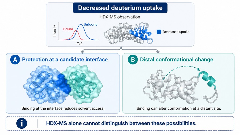
Figure 2. Interpreting HDX-MS Results in Protein-Protein Interaction Studies.
Assessing HDX-MS Evidence and Selecting Follow-Up Methods
The strength of an HDX-MS interface hypothesis depends on sequence coverage, overlap among identified peptides, occupancy of the bound state, replicate agreement, and whether the observed changes exceed technical variability. Sparse coverage around a suspected surface limits the conclusions that can be drawn. Extensive coverage is not sufficient if the relevant changes are isolated or poorly reproducible.
The next method should address the specific question that remains unresolved:
- To assess binding between two proteins: A suitable biophysical assay, such as SPR, BLI, or MST, can characterize their binding behavior under defined in vitro conditions.
- To investigate the contribution of candidate interface residues: Targeted mutagenesis can provide complementary evidence, provided that effects on protein folding and stability are also considered.
- To investigate the spatial arrangement of protein subunits: XL-MS can provide residue-pair distance restraints that complement HDX-MS data. A suitable structural method may be needed when a more detailed complex structure is required.
Frequently Asked Questions
1. Can HDX-MS be used for epitope mapping of a protein-protein pair?
It can locate regions whose exchange changes upon complex formation, which is often useful for epitope or paratope hypothesis generation. Peptide-level protection is still not equivalent to a residue-resolved contact map. Independent binding evidence and, where needed, mutagenesis remain important.
2. Why might two peptides in the same domain show opposite uptake changes?
Binding can bury one local surface while increasing dynamics in an adjacent loop or a remote domain. Opposite changes are therefore biologically possible. They should be interpreted with peptide quality, overlapping coverage, and replicate agreement rather than averaged into a single domain-level conclusion.
3. Does a lack of HDX change mean that two proteins do not bind?
No. Binding may occur without a detectable change in amide exchange, occupancy may be too low, or the affected region may lack peptide coverage. A negative HDX result is a lack of detected uptake difference under the tested conditions, not a disproof of association.
Conclusion
HDX-MS is most informative for protein-protein studies when unbound and bound states are compared under matched labeling conditions and the result is read as a peptide-level conformational map. Protection can support a candidate interface, but remote conformational effects remain an alternative explanation. Researchers with a defined pair and independent binding evidence can consult the MtoZ Biolabs Hydrogen Deuterium Exchange Mass Spectrometry Service for project-specific comparison design.
Reference
- Z. Zhang, D.L. Smith (1993). Determination of amide hydrogen exchange by mass spectrometry: a new tool for protein structure elucidation. Protein Science, 2, 522-531. https://doi.org/10.1002/pro.5560020404
- L. Konermann, J. Pan, Y.H. Liu (2011). Hydrogen exchange mass spectrometry for studying protein structure and dynamics. Chemical Society Reviews, 40, 1224-1234. https://doi.org/10.1039/c0cs00113a
