



Cellular proteomics studies the composition, function, and interactions of proteins within cells. The workflow of cellular proteomics is the ordered set of harvest, extraction, measurement, and interpretation steps used to turn a cell population into protein identifications and, under a separate design, abundance comparisons. In current practice the usual route is bottom-up liquid chromatography-tandem mass spectrometry (LC-MS/MS): proteins are extracted, digested to peptides, separated chromatographically, identified from fragment spectra, and summarized to protein-level tables. Gel electrophoresis, matrix-assisted laser desorption/ionization time-of-flight (MALDI-TOF) intact-mass checks, and antibody assays remain useful in defined niches. They are not interchangeable with a shotgun cellular proteome matrix.
By systematically analyzing cellular proteins, researchers can examine biological processes and disease models, provided the cell state, lysis chemistry, and quantitative claim stay matched. The workflow does not, by itself, inventory every protein a cell can theoretically express, and it does not establish a diagnostic cutoff.
Researchers seeking a broader understanding of cellular protein identification, quantification, and proteomic profiling can explore MtoZ Biolabs' Cellular Proteomics Service
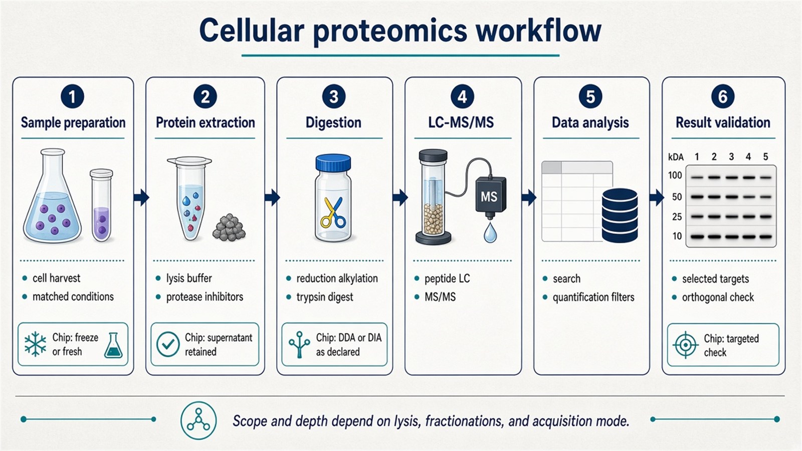
Figure 1. A current cellular proteomics workflow usually proceeds from cell sample preparation and protein extraction through digestion and LC-MS/MS to data analysis and selected result validation; depth still depends on lysis, fractionation, and acquisition mode.
The workflow is a claim-specific sequence, not a single gel, instrument, or software name.
When This Workflow Fits a Cell Study
The workflow fits when the scientific question is protein-resolved at the level of a defined cell population: which proteins are detected after a stated extraction, how the same proteins change across treatments or genotypes, or which candidates should later enter antibody or targeted mass spectrometry confirmation. It is a poor substitute for a bulk colorimetric protein assay, a poor discovery method if only one antibody-defined target is required, and a different experiment from single-cell proteomics or endogenous peptidomics.
|
Study need |
Cellular proteomics workflow often fits |
Reassess before queuing runs |
|---|---|---|
|
Protein inventory of a cell population |
Bottom-up LC-MS/MS after matched lysis and digestion |
Intact-protein catalog or a single Western blot |
|
Relative protein change across cell states |
Label-free or isobaric LC-MS/MS under one QC plan |
Absolute concentration without a matched standard |
|
Gel-based inspection or limited fractionation |
SDS-PAGE or 2-DE as a supporting step |
Treating a gel as a complete proteome |
|
Complex cell lysate identification |
LC-MS/MS database search |
MALDI-TOF peptide-mass fingerprint of an unfractionated lysate |
|
Confirmation of selected proteins |
Western blotting, ELISA, or immunofluorescence |
Using one blot to validate an entire discovery table |
Prefer a gel-free LC-MS/MS workflow when the claim is a cellular protein matrix. Prefer SDS-PAGE or two-dimensional electrophoresis when intact-protein resolution, immunoblotting load control, or limited fractionation is the actual job. Prefer a targeted peptide assay when the protein list is already closed.
Sample Preparation
Sample preparation is the first step in cellular proteomics. It typically involves selecting cell lines, primary cells, or other cell populations that represent the biological state under investigation. Tissue pieces can enter a related proteomics plan, but they are not the same input as a washed cell pellet: extracellular matrix, mixed cell types, and blood contamination change extraction and search assumptions.
Handling should occur on ice or at low temperature to limit protein degradation, and protease inhibitors are commonly included at lysis. Cells are washed to remove residual medium and serum proteins before they are collected as pellets. Adherent cultures require controlled detachment so that residual trypsin does not become an unintended component of the digest. Cells are then disrupted mechanically, by detergent-containing buffers, or by a combination of those approaches to release intracellular proteins. Freeze-thaw cycling of the pellet should be minimized, because repeated thawing changes extractability before any mass spectrometer is started.
Washed cell pellets, frozen cells, lysates, and already extracted protein can all enter a cellular proteomics plan, but the harvest format has to be declared with the contrast. Residual fetal bovine serum and incomplete PBS washes are common sources of non-cellular identifications.
For a broader bottom-up sample-preparation sequence that includes digestion, desalting, and pre-injection handling, researchers can refer to A Comprehensive Workflow for Bottom-Up Proteomics Analysis (with Sample Preparation Techniques) for more detailed guidance.
Protein Extraction
Protein extraction uses an appropriate lysis buffer to solubilize membranes and release proteins. Commonly used buffers contain detergents, and the buffer has to be compatible with the later digestion and labeling chemistry. Strong detergents that help dissolve membrane proteins can suppress digestion or interfere with isobaric labeling if they are not removed. Mechanical disruption, freeze-thaw, and enzymatic cell-wall treatments are adjuncts for difficult cells, not replacements for a declared buffer.
The extract is centrifuged to remove cell debris and insoluble material. The resulting supernatant contains the recovered soluble protein fraction, not the entire theoretical proteome. Insoluble pellets can retain cytoskeletal, nuclear, or membrane proteins if the lysis was too mild. A bulk protein assay such as bicinchoninic acid (BCA) is useful for matching input before digestion. It does not identify which proteins were recovered.
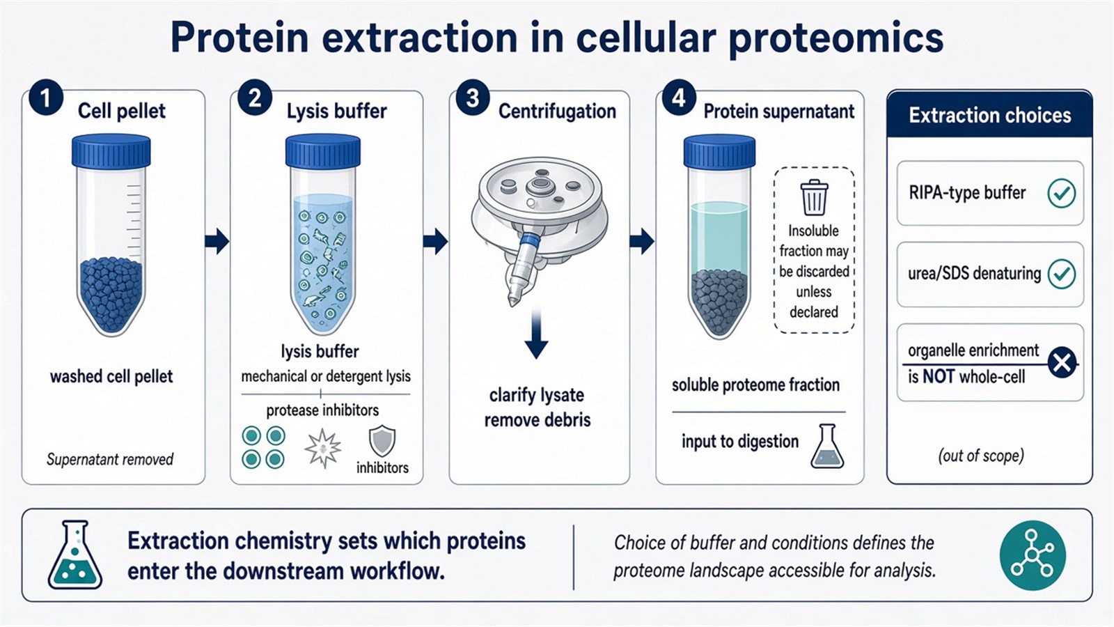
Figure 2. Protein extraction in cellular proteomics typically proceeds from a washed cell pellet through lysis and centrifugation to a soluble supernatant; extraction chemistry sets which proteins enter the downstream workflow.
The supernatant is the protein population that this lysis recovered. It is not proof that unextracted proteins were absent from the cell.
Protein Digestion
Most cellular proteomics identification is bottom-up. Extracted proteins are reduced and alkylated, then digested, most often with trypsin and sometimes with Lys-C, so that peptides rather than intact proteins enter LC-MS/MS. Incomplete digestion, missed cleavage, and detergent carryover reduce identification depth and distort quantitative values. This step is missing from many schematic descriptions that jump from extraction to a gel, yet it is the operational measurement layer behind current shotgun cellular proteomes.
Gel-based workflows can digest proteins in-gel after SDS-PAGE. Gel-free workflows digest in solution or on a bead or filter support, then desalt peptides before injection. The digestion chemistry has to be chosen with the quantification route: amine-reactive tandem mass tags require peptides that can still be labeled after cleanup.
Protein Separation
Protein separation in classical cellular proteomics often used electrophoresis. Sodium dodecyl sulfate polyacrylamide gel electrophoresis (SDS-PAGE) separates proteins mainly by molecular weight. Two-dimensional electrophoresis (2-DE) adds an isoelectric-point dimension and can increase resolution for intact proteins. Those gels remain useful for visual inspection, immunoblotting, intact-mass work, and limited fractionation.
Discovery LC-MS/MS studies more often skip a protein gel and separate peptides by reversed-phase liquid chromatography after digestion. That gel-free route is the usual path to a cellular protein identification table. A gel is not required for every cellular proteomics project, and running SDS-PAGE does not by itself produce a proteome-wide matrix.
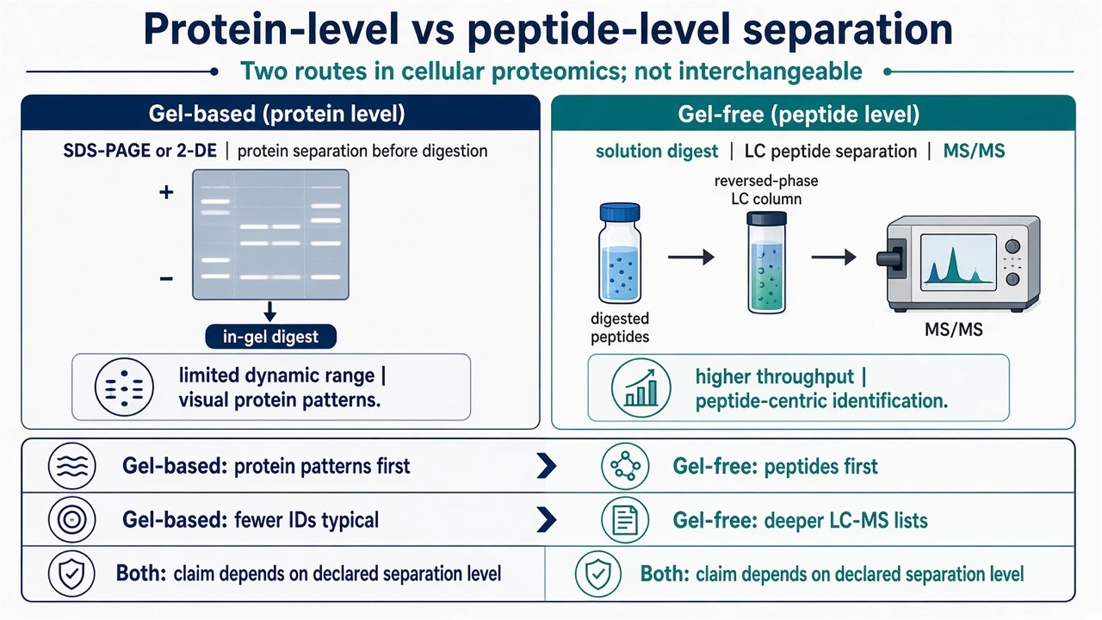
Figure 3. Protein separation in cellular proteomics may use SDS-PAGE or 2-DE at the protein level, or gel-free LC-MS/MS at the peptide level; the two routes answer different claims and are not interchangeable.
Protein Identification
Protein identification is achieved through mass spectrometry. MALDI-TOF can report intact mass or a peptide-mass fingerprint for relatively simple samples. LC-MS/MS is the workhorse for complex cell lysates: liquid chromatography separates peptides, tandem mass spectrometry records fragment ions, and software matches those spectra to a protein database. Sequence-bearing fragment evidence, not a single intact mass, is what usually supports identity in a cellular extract.
Database choice has to match the species and the expected contaminants. A human cell line search that omits bovine serum proteins can mis-assign residual medium proteins. Reverse or decoy sequences are used to control false identifications. Protein inference then summarizes peptide evidence to protein groups. Shared peptides and one-hit identifications need declared filters before the table is read as a cellular inventory.
Protein Quantification
Protein quantification is the step that estimates expression differences of the same proteins across cell states. Common routes include labeling and label-free methods. Labeling methods such as tandem mass tags (TMT) and isobaric tags for relative and absolute quantitation (iTRAQ) allow multiple samples to be compared after mixing. Label-free methods, including data-dependent peak-area quantification and data-independent acquisition such as SWATH-MS, compare independent injections of unlabeled digests (Thompson et al. 2003; Gillet et al. 2012; Cox et al. 2014).
These routes are not interchangeable names for one instrument method. TMT or iTRAQ fit a closed sample set that can share one plex, at the cost of plex design and possible ratio compression. Label-free LC-MS/MS fits open enrollment and later-arriving samples, at the cost of run-to-run normalization. SWATH-style acquisition is a fragment-window strategy inside the unlabeled family. It does not, by itself, quantify the entire proteome or report absolute concentration. Amount claims need matched internal standards and a calibration model.
When sample number, multiplexing, or acquisition strategy makes the choice less straightforward, researchers can refer to How to Choose a Quantitative Proteomics Strategy for a more detailed strategy-selection framework.
Data Analysis
Mass spectrometry data require computational and statistical processing before they become a protein table. Software tools such as MaxQuant and Proteome Discoverer are widely used to assign peptide-spectrum matches, apply false-discovery filters, and summarize quantitative values (Cox et al. 2014). Data-independent datasets are often processed in dedicated tools rather than in a DDA-centric pipeline. The analysis includes protein identification, quantification, and a first pass of annotation, typically organized as tables for later visualization.
A larger intensity means more ion current was recorded for that feature under the stated digestion, loading, and acquisition plan. Missing values, shared peptides, and unmatched batches remain part of the record. Functional annotation at this stage is a mapping of identifiers. It is not yet evidence that a pathway was biologically activated.
Bioinformatics Analysis
After the protein table is closed, bioinformatics analysis interprets functions, pathways, and interaction networks to place the results in a biological context. Online databases and software such as STRING and DAVID are commonly used for enrichment analysis and network construction. Those maps can highlight candidate biomarkers or therapeutic hypotheses for follow-up. Enrichment reports overrepresentation relative to a defined background. It does not prove pathway activity, and a STRING edge is not an experimentally confirmed interaction in that cell line.
GO and KEGG-style summaries inherit every bias of the quantified proteome, including lysis recovery and identification filters. Hypothesis generation is a valid use of this step. Treating an enrichment plot as a mechanism is not.
Result Validation
Experimental methods are then used to validate selected results from the discovery table. Western blotting, enzyme-linked immunosorbent assay (ELISA), or immunofluorescence staining can test whether nominated proteins behave as expected in the same cellular system. Those assays confirm the proteins they were designed to see. They do not re-measure the rest of the proteome, and a failed antibody assay can reflect epitope or linearity problems rather than a false mass spectrometry identification.
Orthogonal confirmation belongs on proteins that will carry the biological claim. Re-running the same LC-MS/MS search is reproducibility review, not independent validation.
Experimental Limits and Design Boundaries
Post-harvest proteolysis, residual serum, keratin, and unmatched lysis change which proteins appear. Detergent and nucleic-acid carryover disturb digestion and chromatography. Gel-only workflows under-sample complex lysates. Unlabeled batches drift without run-order control. Isobaric plexes cannot quietly absorb samples that arrive after mixing. Bioinformatics enrichment cannot rescue a poorly matched harvest.
Cellular proteomics of washed pellets is not single-cell proteomics, not HLA immunopeptidomics, and not endogenous peptidomics without digestion. Diagnostic cutoffs, companion-diagnostic claims, and absolute copy-number statements sit outside ordinary discovery reporting unless a separately validated assay framework is in place.
Once the cell state, extraction chemistry, and quantitative claim are defined, the project can be scoped as a cellular proteomics study. Researchers can review the MtoZ Biolabs Cellular Proteomics Service for project-specific sample evaluation, feasibility assessment, and workflow planning.
Frequently Asked Questions
1. Is SDS-PAGE required in every cellular proteomics workflow?
No. SDS-PAGE and 2-DE remain useful for intact-protein inspection or limited fractionation. Gel-free peptide LC-MS/MS is the usual route to a cellular protein identification table.
2. Can MALDI-TOF replace LC-MS/MS for a complex cell lysate?
Not as a default. MALDI-TOF is useful for intact mass or simple peptide-mass fingerprints. Complex cellular extracts generally need LC-MS/MS fragment evidence and database search.
3. Are TMT, iTRAQ, and SWATH-MS interchangeable?
No. TMT and iTRAQ are isobaric labeling chemistries for mixed-sample comparison. SWATH-MS is a data-independent, usually unlabeled, acquisition mode. The chemistry and the acquisition mode answer different design constraints.
4. Does pathway enrichment prove that a biological pathway was activated?
No. Enrichment tests whether identified or differential proteins are overrepresented relative to a background. It is a hypothesis-generating summary, not a direct activity assay.
5. Does one Western blot validate the whole cellular proteome table?
No. Antibody assays confirm selected proteins under their own linearity and epitope limits. They do not re-measure proteins that were not targeted.
Reference
- Aebersold R., Mann M. (2016). Mass-spectrometric exploration of proteome structure and function. Nature, 537, 347-355. https://doi.org/10.1038/nature19949
- Cox J., Hein M.Y., Luber C.A., et al. (2014). Accurate proteome-wide label-free quantification by delayed normalization and maximal peptide ratio extraction, termed MaxLFQ. Mol. Cell. Proteomics, 13, 2513-2526. https://doi.org/10.1074/mcp.M113.031591
- Gillet L.C., Navarro P., Tate S., et al. (2012). Targeted data extraction of the MS/MS spectra generated by data-independent acquisition: a new concept for consistent and accurate proteome analysis. Mol. Cell. Proteomics, 11, O111.016717. https://doi.org/10.1074/mcp.M111.016717
- Thompson A., Schafer J., Kuhn K., et al. (2003). Tandem mass tags: a novel quantification strategy for comparative analysis of complex protein mixtures by MS/MS. Anal. Chem., 75, 1895-1904. https://doi.org/10.1021/ac0262560
