



Whole cell proteomics is the proteomic analysis of an unfractionated cell population, usually as a whole-cell lysate rather than as isolated organelles. Cells rely on proteins for structure, signaling, metabolism, and stress responses, so examining composition, function, and coordinated abundance changes can clarify physiology and disease models. The usual current route is bottom-up liquid chromatography-tandem mass spectrometry (LC-MS/MS) of a digested extract. That measurement reports the protein complement recovered under a stated lysis and acquisition plan. It does not inventory every protein a cell can theoretically express, and it does not assign those proteins to a compartment.
Protein microarray and two-dimensional gel electrophoresis (2-DE) remain useful in defined niches. They are not interchangeable with a shotgun whole-cell protein matrix. Clinical-style claims, including diagnostic cutoffs and individualized treatment decisions, sit outside ordinary research reporting unless a separately validated assay framework is in place.
Researchers who already have defined samples, comparison groups, or a specific project objective can also review the MtoZ Biolabs Cellular Proteomics Service for project-specific feasibility and analysis planning.
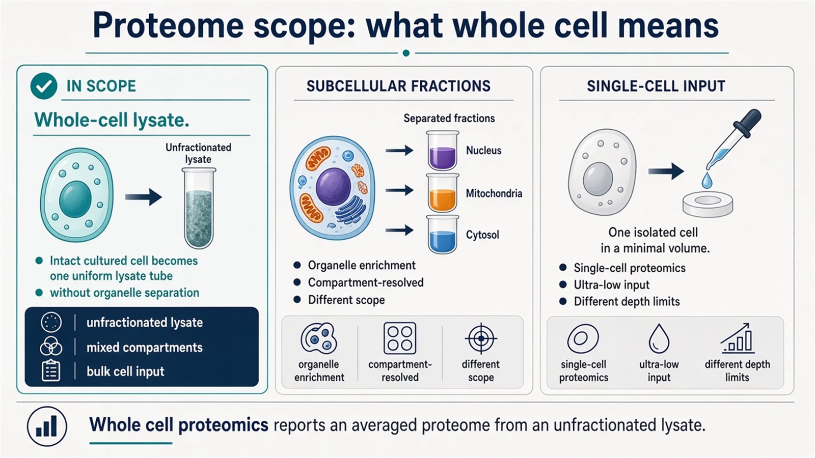
Figure 1. Whole cell proteomics measures an unfractionated whole-cell lysate; it is a different scope from subcellular fractions and from single-cell input.
Whole cell proteomics is defined by the absence of organelle enrichment, not by a promise to see every protein.
When Whole Cell Proteomics Fits
The approach fits when the scientific question is the detectable proteome of a defined cell state after matched harvest and lysis: treatment versus control, genotype contrasts, differentiation time points, or cancer-cell models used to prioritize protein changes. It is a poor substitute for organelle localization, a poor substitute for a single-antibody Western blot, and a different experiment from single-cell proteomics.
|
Study need |
Whole cell proteomics often fits |
Reassess before queuing runs |
|---|---|---|
|
Unfractionated cell-state comparison |
LC-MS/MS of a whole-cell lysate |
The claim requires nuclear, mitochondrial, or membrane identity |
|
Open protein inventory from cultured cells |
Gel-free shotgun identification and relative quantification |
One named protein with a validated antibody |
|
Organelle or compartment composition |
Subcellular fractionation first |
Treating a whole-cell table as localization evidence |
|
Known-target screening at scale |
Protein microarray when probes exist |
Discovery of unexpected proteins not on the array |
|
Intact-protein resolution on a gel |
2-DE as a supporting or historical method |
Using 2-DE as a complete modern whole-cell matrix |
Prefer whole cell proteomics when the contrast lives in the unfractionated extract. Prefer subcellular proteomics when the claim is compartment composition or translocation. Prefer single-cell proteomics only when heterogeneity of individual cells is the object, not when a washed pellet of millions of cells is available.
When the scientific question is intracellular localization rather than an unfractionated inventory, researchers can refer to Guide to Subcellular Proteomics: From Basic Concepts to Applications for a more detailed explanation of fractionation-based design.
Analytical Principles Behind a Whole-Cell Protein Table
A whole-cell protein table is produced by a chain of decisions: extraction chemistry, separation level, identification evidence, and, when required, a quantitative design. Naming those steps prevents treating “cellular proteomics principles” as a generic background summary detached from the unfractionated claim.
1. Protein extraction and sample integrity
Proteins are released from washed cells with a lysis buffer that typically combines detergent, salt, buffering agents, and protease inhibitors so that membranes break and proteins remain soluble enough for digestion. Matched harvest, residual medium removal, and lysis completeness decide which proteins enter the digest. Incomplete lysis, unmatched detergent strength, or serum carryover can change the recovered inventory more than a later search-parameter tweak.
2. Separation before or after digestion
After extraction, interfering material must be reduced and the proteome presented to the mass spectrometer at a workable complexity. Protein-level routes include SDS-PAGE or 2-DE, which separate intact proteins by size and charge before in-gel digestion. Peptide-level gel-free routes digest the lysate in solution and separate peptides by liquid chromatography, usually reversed-phase nanoLC, immediately before MS/MS. Chromatography based on polarity or hydrophobicity is therefore most often a peptide step in modern whole-cell work, not a substitute for declaring whether the experiment started as a protein gel or a solution digest.
3. Identification by tandem mass spectrometry
Bottom-up identification digests proteins, most often with trypsin, into peptides. The mass spectrometer records precursor m/z values and fragment spectra. Spectra are matched to a searchable database such as UniProt or NCBI under declared enzyme, mass-tolerance, and false-discovery rules. Peptide-spectrum matches are then inferred to protein groups. A single weak match is limited evidence; protein reporting usually requires uniqueness and score filters stated in the project plan.
4. Relative quantification across cell states
Quantitative analysis is a separate claim from identification. Common relative strategies include isobaric labeling such as tandem mass tags (TMT) or iTRAQ, metabolic labeling such as stable isotope labeling by amino acids in cell culture (SILAC) when cells can be fully labeled, and unlabeled methods based on precursor peak areas or data-independent acquisition fragment chromatograms. Labeling supports multiplexed comparison inside a designed channel set. Label-free routes keep samples as independent runs and rely on alignment and normalization. Absolute amount still needs matched standards. SILAC is a labeling strategy, not a label-free method.
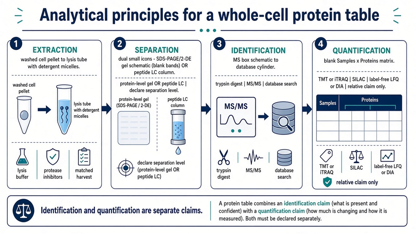
Figure 2. A whole-cell protein table depends on declared extraction, separation level, tandem MS identification, and, when needed, a relative quantification design.
Identification names proteins recovered from the lysate. Quantification compares the same protein across designed cell states.
When sample number, multiplexing, or acquisition strategy makes the choice less straightforward, researchers can refer to DIA vs. DDA in Label-Free Quantitative Proteomics: A Comparative Analysis for a more detailed strategy-selection framework.
Methodologies Used for Whole-Cell Inventories
1. Liquid Chromatography-Mass Spectrometry (LC-MS/MS)
LC-MS/MS merges chromatographic peptide separation with tandem mass spectrometry and is the workhorse for current whole cell proteomics. Proteins are extracted from the unfractionated lysate, digested, separated, identified from fragment spectra, and, under a separate design, quantified (Aebersold and Mann 2016). Label-free peak-area methods, data-independent acquisition, and isobaric tags can all operate on this platform. The method reports peptide-supported protein groups recovered from that lysis. Membrane proteins, extreme pI species, and low-abundance regulators can still be missing if extraction, digestion, or acquisition do not capture them.
2. Protein Microarray Technology
Protein microarrays can simultaneously detect and evaluate large numbers of proteins by immobilizing proteins or antibodies on a chip so that sample proteins bind to those probes. The format is effective for large-scale screening and differential analysis of targets that are already represented on the array. It is not an open whole-cell discovery method: proteins absent from the probe set remain invisible, and binding still depends on epitope access and reagent quality. Use an array when the target list is closed. Use LC-MS/MS when the protein list is still open.
3. Two-Dimensional Gel Electrophoresis (2-DE)
2-DE uses isoelectric focusing and SDS-PAGE to separate intact proteins from complex samples. It can offer high resolution for visualized proteoforms and remains useful for limited fractionation or immunoblotting follow-up. It faces well-known limits with very large or hydrophobic proteins, low-abundance species, and highly complex lysates. In current whole cell proteomics, 2-DE is a supporting or historical route, not the default path to a deep protein identification table.
From Unfractionated Lysis to a Protein Matrix
A practical LC-MS/MS whole cell proteomics sequence starts with cell harvest, proceeds through unfractionated lysis without organelle enrichment, then digestion and peptide cleanup, and ends in LC-MS/MS identification and a declared quantitative comparison. Harvest matching, residual medium, and lysis chemistry decide which proteins enter the digest. Organelle pellets that are discarded or never generated cannot be reconstructed later by bioinformatics localization prediction.
Filter-aided or in-solution digestion methods are commonly used to move a detergent-containing whole-cell extract into a peptide sample suitable for nanoLC-MS/MS (Wiśniewski et al. 2009). Quantification must be named with the method: relative change of the same protein across cell states, or amount only when standards and calibration are present. Dynamic proteomic states under different conditions require designed time points or treatments. They are not recovered by analyzing one untreated pellet more deeply.
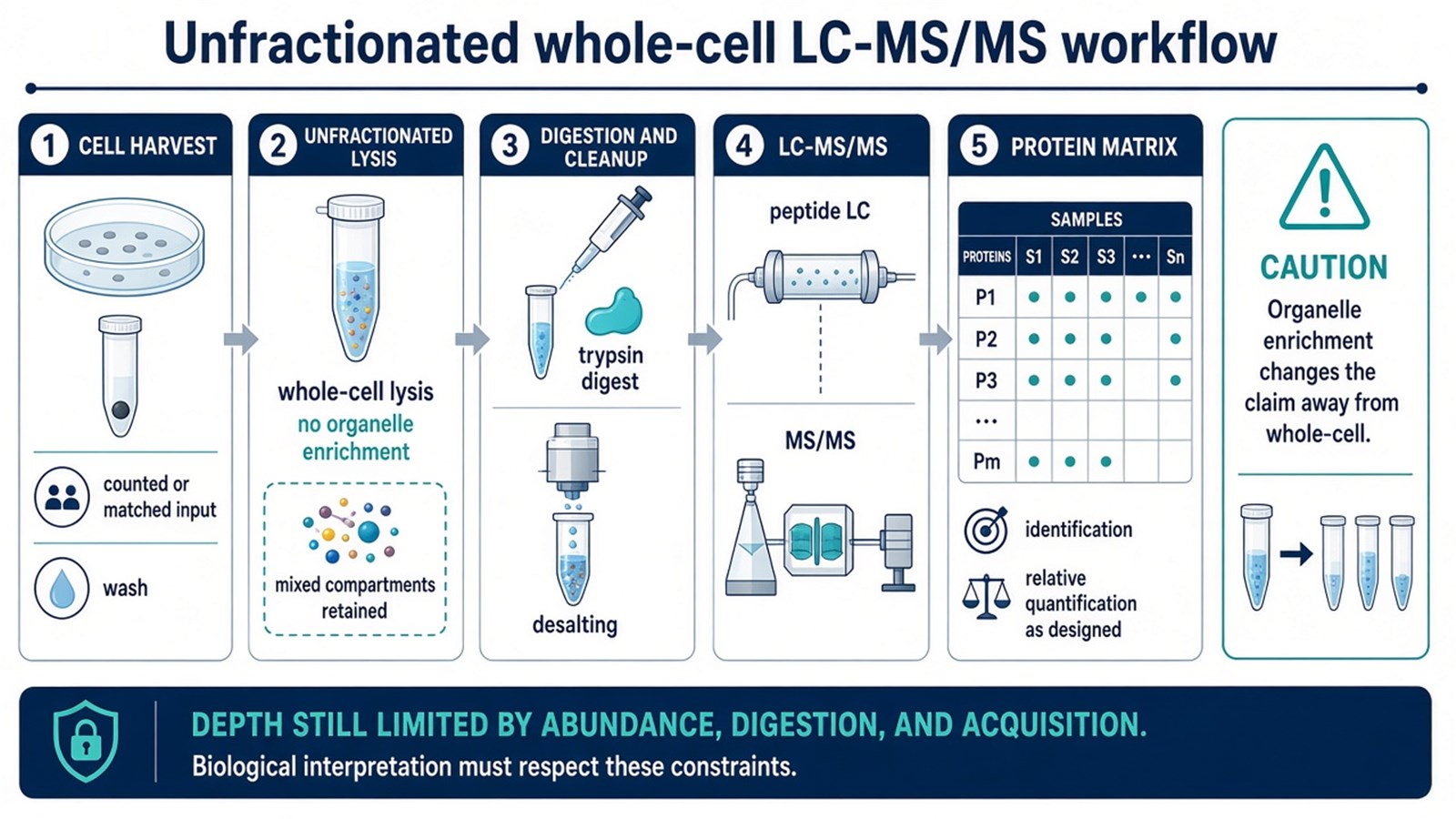
Figure 3. Whole cell proteomics typically proceeds from cell harvest through unfractionated lysis and digestion to LC-MS/MS, without organelle enrichment.
Comparability is built at harvest and unfractionated lysis. Search software cannot restore proteins that were never extracted.
Cell pellets, tissues, and biofluids are not interchangeable whole-cell inputs. Tissue homogenates mix cell types; plasma is not a cell lysate. For sample-matrix differences that change preparation, researchers can refer to Proteomics Service of Different Sample Types for more detailed guidance.
Protein Interactions Sit Outside a Whole-Cell Inventory
Proteins often act in complexes, and interaction mapping can use yeast two-hybrid, co-immunoprecipitation, or pull-down enrichment followed by mass spectrometry. Those designs answer which partners associate with a bait under enrichment conditions. A whole-cell lysate inventory lists proteins recovered without bait enrichment and does not, by itself, prove physical interaction, complex membership, or pathway activity. When the scientific object is an interactome rather than an unfractionated cell-state matrix, the experiment should be scoped as interaction proteomics from the start.
Research Applications
Beyond foundational research on cellular physiology and pathology, whole cell proteomics is used to generate protein-level hypotheses in disease models, compound treatment, and translational research. Comparing whole-cell proteomes in cancer-cell or other disease models can highlight proteins associated with progression or drug response for later validation. In drug-discovery models, the same design can support target hypothesis generation, mechanism-of-action surveys, and off-target protein changes when matched vehicle controls exist. In precision-medicine research, a model or donor proteomic profile can stratify experimental groups. Those lists are candidate maps. They are not diagnostic tests, approved companion diagnostics, or personalized prescriptions.
Advantages and Challenges of Whole Cell Proteomics
1. Advantages
The systematic nature of whole cell proteomics provides a cell-level protein inventory under one lysis and acquisition plan, which can reveal coordinated abundance changes and support later network or pathway hypotheses. It can also surface disease-associated proteins in cell models without requiring an antibody for each name. That panoramic view is panoramic relative to the recovered extract, not relative to the theoretical proteome.
2. Challenges
Data analysis is computationally demanding and requires declared identification filters, normalization, and experimental design. Capturing dynamic proteomic states under varying conditions remains difficult: one snapshot does not describe turnover, translocation, or modification-level regulation. Low-abundance and hydrophobic proteins are still under-sampled in many unfractionated lysates. Enrichment, subcellular fractionation, or a targeted assay is then a different experiment, not a software setting.
Experimental Limits and Design Boundaries
Whole cell proteomics does not report where a protein sits, which cell in a mixed culture produced it, or whether a missing identification means biological absence. Residual serum, keratin, unmatched lysis, and single-peptide identifications can overstate the cellular proteome. Pathway enrichment from a differential list is overrepresentation against a background, not proof of pathway activity. Interaction claims require enrichment designs. Localization claims require fractionation or imaging.
Diagnostic cutoffs, companion-diagnostic claims, and guaranteed improvements in therapeutic success sit outside ordinary research whole cell proteomics. Once the cell state, unfractionated lysis plan, and quantitative claim are defined, the project can be scoped. Researchers can review the MtoZ Biolabs Cellular Proteomics Service for project-specific sample evaluation, feasibility assessment, and workflow planning.
Frequently Asked Questions
1. Does whole cell proteomics identify all proteins in a cell?
No. It identifies proteins recovered from an unfractionated lysate under a stated extraction, digestion, and search plan. Missing identifications are not proof of biological absence.
2. Is whole cell proteomics the same as subcellular proteomics?
No. Whole cell proteomics skips organelle enrichment. Subcellular proteomics requires fractionation or isolation before LC-MS/MS and is the route when compartment identity is the claim.
3. Is whole cell proteomics the same as a generic cellular proteomics principle page?
No. Principles of extraction, separation, identification, and quantification apply only after the sample scope is fixed as an unfractionated whole-cell lysate. Organelle enrichment, bait interactomes, and single-cell inputs change the claim.
4. Is 2-DE still required for whole cell proteomics?
No. 2-DE remains useful for intact-protein resolution or limited fractionation. Gel-free LC-MS/MS is the usual current path to a whole-cell identification table.
5. Can a protein microarray replace shotgun LC-MS/MS?
Not for an open protein list. Arrays measure targets represented by their probes. Unexpected proteins require a discovery method such as LC-MS/MS.
6. Can whole cell proteomics be read as a clinical diagnostic result?
Not by default. Research tables can prioritize biomarker or target candidates. Diagnostic or treatment decisions need a separately validated assay framework.
Reference
- Aebersold R., Mann M. (2016). Mass-spectrometric exploration of proteome structure and function. Nature, 537, 347-355. https://doi.org/10.1038/nature19949
- Wiśniewski J.R., Zougman A., Nagaraj N., Mann M. (2009). Universal sample preparation method for proteome analysis. Nat. Methods, 6, 359-362. https://doi.org/10.1038/nmeth.1322
- Yates J.R., Gilchrist A., Howell K.E., Bergeron J.J. (2005). Proteomics of organelles and large cellular structures. Nat. Rev. Mol. Cell Biol., 6, 702-714. https://doi.org/10.1038/nrm1711
