



Ligand-receptor analysis tests binding between a defined ligand and a defined receptor. It is not a single method. The ligand may be a small molecule, a peptide, or a protein, and the receptor may be a membrane protein, a nuclear receptor, or a soluble receptor ectodomain. Transmembrane proteins are a common receptor class, not the only one. The result reports binding for that pair. It does not, by itself, explain cellular communication or identify a safe drug candidate.
Hormones, neurotransmitters, and drugs are common biological contexts for these binding studies. Where a receptor construct and a ligand are already defined, the MtoZ Biolabs Ligand Receptor Analysis Service can be used to check feasibility and choose a binding format.
How Ligand-Receptor Analysis Differs from a Protein–Ligand Interaction Profile
A protein–ligand interaction profile records the measurement for a defined pair, such as a sensorgram, a thermogram, a docked pose, or a Förster resonance energy transfer (FRET) trace. Ligand-receptor analysis asks whether the protein is being studied as a receptor and which binding event that measurement reports. Those measurement types are set out on the Protein Ligand Interaction Profile page.
Single-cell ligand–receptor analysis is a different experiment. An inferred pair from transcript counts is not a binding measurement. Expression of a ligand and a receptor in neighboring cells does not report occupancy, affinity, or a site.
Native mass spectrometry (native MS) can report intact-mass evidence that the ligand remains bound when the complex survives ionization. It does not replace a kinetic assay or a cell-based occupancy assay.
Sample Preparation Is a Construct Choice
Ligand-receptor analysis does not typically extract both partners from cells, tissues, or whole organisms. The receptor is often a recombinant ectodomain, a membrane vesicle, a solubilized receptor, or a cell that still displays the receptor. The ligand is often synthetic or recombinant. Purity and a functional check matter for the format that will be run. They do not directly determine the accuracy of the results.
Affinity chromatography and gel filtration can polish a soluble construct. A detergent-solubilized membrane receptor is handled differently from a soluble globular ligand. For a membrane protein, the lipid or detergent system needs to be compatible with the instrument.
What a Binding Measurement Reports
Mass spectrometry, FRET, and surface plasmon resonance (SPR) are separate measurements. Affinity, kinetics, and a binding site are not three outputs of every one of these methods.
SPR can record association and dissociation of an immobilized receptor or ligand. Real-time means the sensorgram is collected during the injection. FRET reports energy transfer between two labels. Mass spectrometry reports a mass, a selected ligand, or a covalent peptide, depending on the first event. None of those files is a precise binding site unless a separately validated structure or a localized adduct is already named.
How mass spectrometry, FRET, and SPR report different binding events is discussed on the How to Detect Protein and Small Molecule Interactions page.
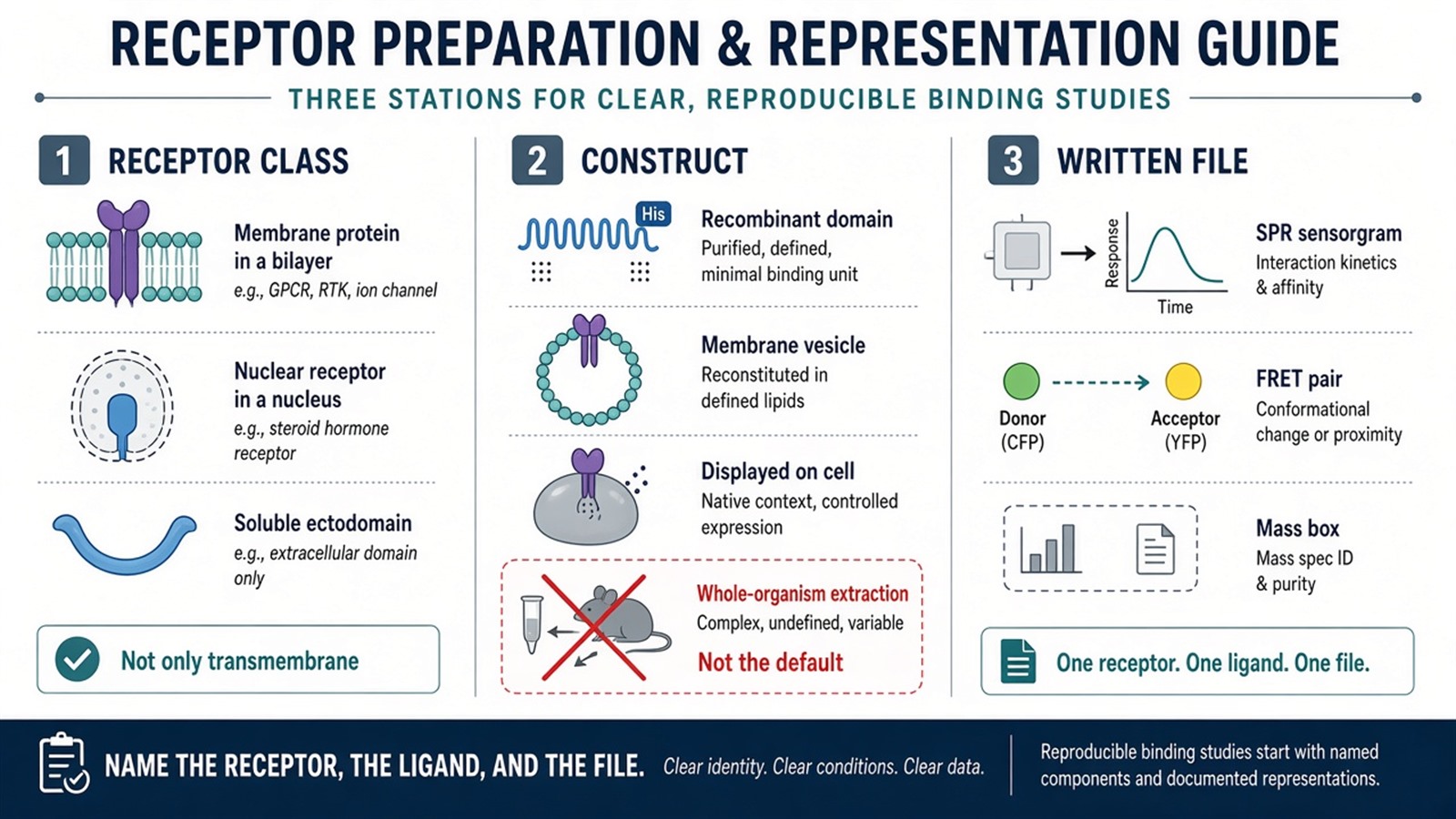
Figure 1. The receptor class and the construct are defined before the measurement, which may be a sensorgram, a label trace, or a mass.
The receptor, the ligand, and the measurement should be identified before a result is used to support a drug-target conclusion.
Sensitivity, Specificity, and Kinetics Depend on the Assay
Sensitivity at low ligand concentration depends on the assay window. Selectivity among ligands or receptor subtypes comes from a comparison across those ligands or subtypes. Real-time kinetic data come from formats that record a time course, chiefly SPR or a related optical biosensor.
SPR and calorimetry require a pure and stable sample. An in-vitro binding result describes the system that was measured. A second method can add another in-vitro measurement, but it does not reproduce the cellular environment.
Proliferation, differentiation, and apoptosis are measured in cell assays. A binding result does not report those cellular responses, and it does not by itself support a diagnostic or treatment conclusion.
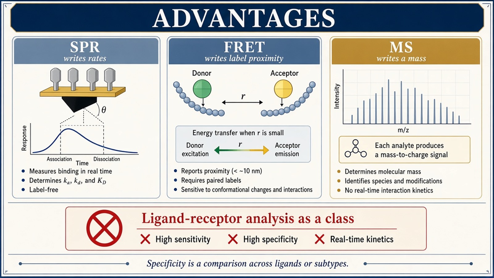
Figure 2. SPR reports binding rates, FRET reports label proximity, and MS reports a mass.
Experimental Limits
The next experiment follows the readout already named: a kinetic sensorgram, cell-based occupancy, or intact-mass evidence from native MS.
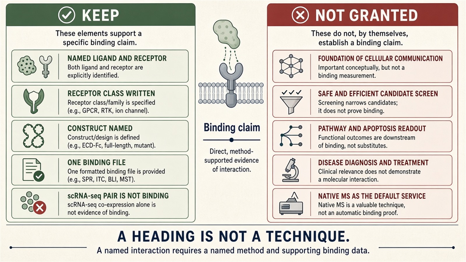
Figure 3. A ligand-receptor result is tied to one receptor, one ligand, and one measurement.
Project Planning and Service Scope
A project request should name the receptor class (membrane, nuclear, or soluble), the construct, whether cells, vesicles, or purified protein will be used, and which readout is required after a positive result. MtoZ Biolabs can run the work as SPR, a ligand-binding assay, or mass spectrometry when that format matches the question. The result is then interpreted according to that format.
For a receptor ligand-binding assay, the MtoZ Biolabs Receptor Ligand Binding Assay Service covers sample evaluation, feasibility assessment, and workflow planning.
Frequently Asked Questions
1. Is ligand-receptor analysis one technique?
No. It is a binding question answered by a named measurement, such as a sensorgram, a label trace, or a mass.
2. Are receptors predominantly transmembrane proteins?
Many receptors are transmembrane proteins. Nuclear receptors and soluble receptors are also receptors.
3. Does the workflow start by extracting both partners from an organism?
Not as a default. The receptor is often a construct. The ligand is often synthetic.
4. Do MS, FRET, and SPR provide sites as well as affinity?
SPR can report association and dissociation rates. FRET reports proximity between labels. A binding site requires a separate structural or localization result.
5. Is Native MS the related service for this heading?
Only when the question is whether the intact complex can be observed by mass. A sensorgram or a cell-based occupancy assay is a different measurement.
Reference
-
Hulme E.C., Trevethick M.A. (2010). Ligand binding assays at equilibrium: validation and interpretation. Br. J. Pharmacol., 161, 1219-1237. https://doi.org/10.1111/j.1476-5381.2010.00831.x
-
Renaud J.P., Chung C.W., Danielson U.H., et al. (2016). Biophysics in drug discovery: impact, challenges and opportunities. Nat. Rev. Drug Discov., 15, 679-698. https://doi.org/10.1038/nrd.2016.123
-
Rosenbaum D.M., Rasmussen S.G.F., Kobilka B.K. (2009). The structure and function of G-protein-coupled receptors. Nature, 459, 356-363. https://doi.org/10.1038/nature08144
-
Rich R.L., Myszka D.G. (2000). Advances in surface plasmon resonance biosensor analysis. Curr. Opin. Biotechnol., 11, 54-61. https://doi.org/10.1016/S0958-1669(99)00054-3
