



Plasma proteome profiling is the LC-MS/MS study of proteins circulating in plasma, including which proteins are detected, how their abundance changes, and, under a separate design, selected post-translational modifications. Plasma is a convenient biofluid because it can be sampled repeatedly and contains proteins secreted by the liver, proteins leaked or shed from tissues, and proteins of the coagulation and immune systems. It is not a uniform mixture of proteins from every organ in equal measure. Albumin, immunoglobulins, and a small set of other high-abundance proteins dominate the measurable signal, so a profiling experiment reports the proteome recovered after a stated collection, depletion or enrichment choice, digestion, and acquisition plan (Anderson and Anderson 2002; Geyer et al. 2017).
That matrix can support research on physiological state, disease models, treatment response, and candidate biomarker generation. It does not, by itself, provide an early-diagnosis test, a personalized prescription, or a companion-diagnostic cutoff.
Researchers who already have defined samples, comparison groups, or a specific project objective can also review the MtoZ Biolabs Plasma Proteomics Analysis Service for project-specific feasibility and analysis planning.
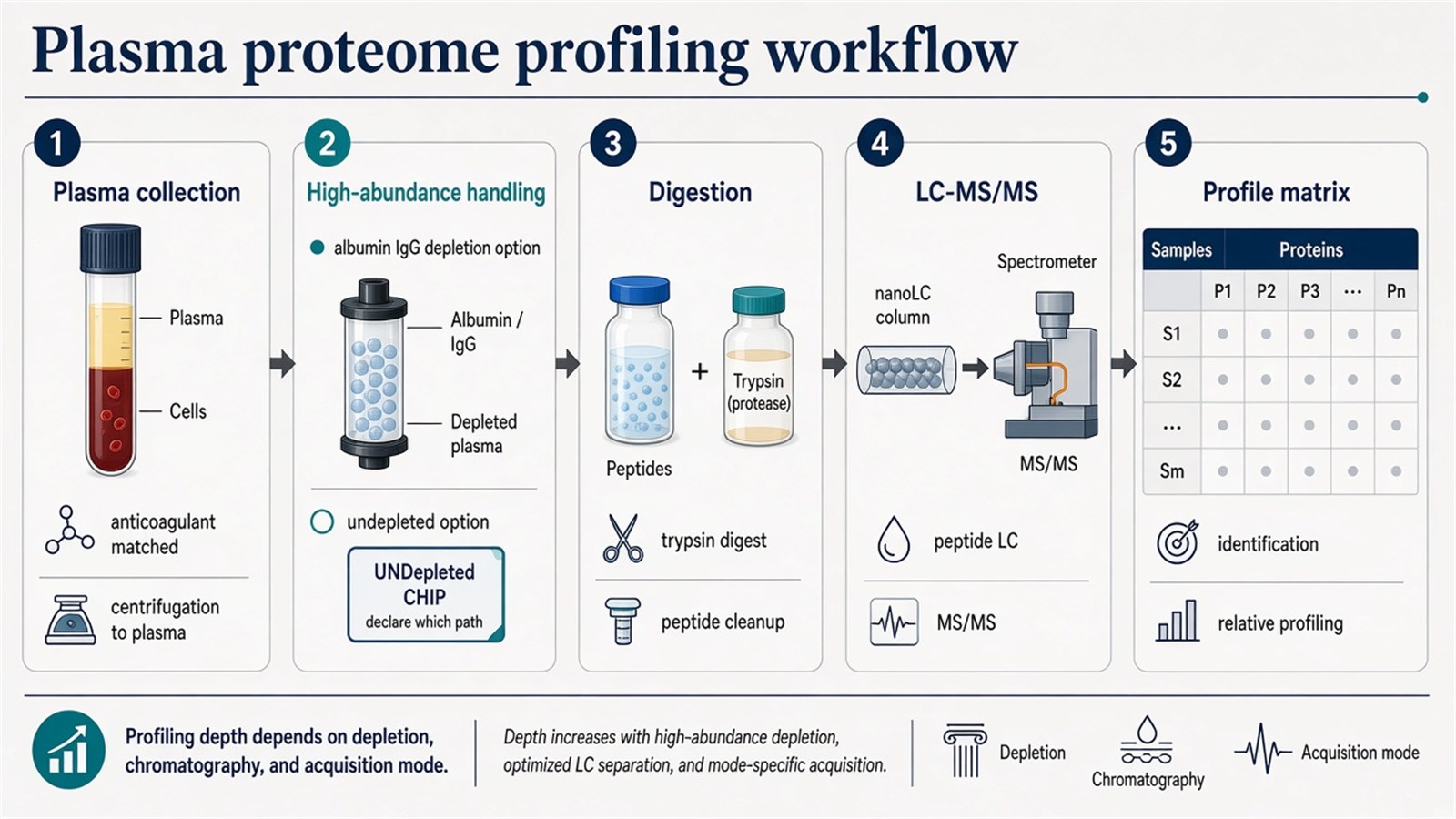
Figure 1. Plasma proteome profiling typically proceeds from matched plasma collection through high-abundance handling and digestion to LC-MS/MS; depth depends on depletion, chromatography, and acquisition mode.
A plasma protein table is only as comparable as the anticoagulant, freeze-thaw history, and high-abundance handling used to produce it.
When Plasma Proteome Profiling Fits
The experiment fits when the scientific claim is protein-resolved in plasma: which proteins are detected after a declared preparation, how the same proteins change across groups or time points, or which candidates should later enter a targeted assay. It is a poor substitute for a tissue proteome, a poor discovery method if only one ELISA antigen is required, and a different matrix from serum.
|
Study need |
Plasma proteome profiling often fits |
Reassess before queuing runs |
|---|---|---|
|
Open plasma protein inventory |
Undepleted or depleted LC-MS/MS under one QC plan |
A single named antigen with a validated immunoassay |
|
Group or pre- versus post-treatment comparison |
Label-free, DIA, or TMT profiling on matched plasma |
Mixing EDTA plasma with serum in one model |
|
Lower-abundance candidate discovery |
Immunoaffinity depletion, filtration, or other enrichment |
Treating depletion as complete recovery of tissue leakage proteins |
|
Coagulation-related proteins |
Consistently collected plasma |
Serum collected after clotting |
|
Diagnostic cutoff or treatment decision |
A validated clinical assay framework |
Reading a discovery table as a diagnostic result |
Prefer plasma when coagulation proteins must remain in the sample and the anticoagulant can be held constant, typically EDTA or citrate rather than heparin. Prefer serum only when the study was designed for the clotted matrix. Prefer a targeted peptide assay when the protein list is already closed.
Plasma, serum, and cerebrospinal fluid are related biofluids with different collection chemistry and protein backgrounds. For matrix-specific planning across those fluids, researchers can refer to Serum / Plasma / CSF Proteomics Service for more detailed project scoping.
High-Abundance Proteins and Sample Pretreatment
The process of plasma proteome profiling runs from sample pretreatment through digestion, LC-MS/MS, and data interpretation. During processing, high-abundance proteins can overshadow lower-abundance ones and complicate their detection. Immunoaffinity depletion and filtration are therefore used in many designs to remove high-abundance proteins and enrich remaining targets, which can improve detection of a deeper slice of the plasma proteome. Depletion also removes proteins bound to albumin or other carriers and can change quantitative ratios. Undepleted workflows, including some DIA designs, keep the native abundance structure at the cost of shallower access to lower-abundance species. High-abundance handling is a declared experimental choice, not a hidden software correction.
Hemolysis, repeated freeze-thaw, and unmatched collection tubes create protein changes that later look like biology. Samples with visible hemolysis or precipitation should be flagged before they enter a cohort comparison.
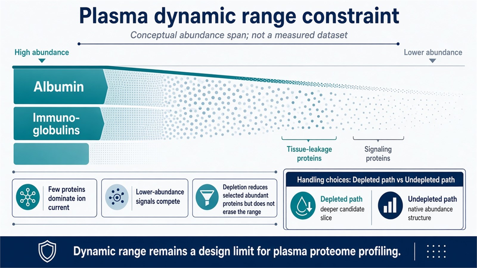
Figure 2. Plasma proteome profiling is constrained by dynamic range: a few high-abundance proteins can overshadow lower-abundance proteins unless handling is declared, and depletion does not erase the range.
Depletion can reveal lower-abundance peptides. It does not convert a plasma table into a tissue proteome.
Digestion, LC-MS/MS, and Acquisition
After pretreatment, proteins are hydrolyzed into peptides using enzymes such as trypsin, with reduction and alkylation as needed, so that peptides rather than intact plasma proteins enter the mass spectrometer. High-resolution LC-MS/MS then supports qualitative assignment and, under a quantitative design, comparison of peptide signals across samples.
Data-dependent acquisition (DDA) selects precursors for fragmentation and remains useful for identification-centric or library-building runs, at the cost of stochastic missingness in complex plasma. Data-independent acquisition (DIA) fragments scheduled windows and is often used when consistent quantification across a plasma cohort is the priority (Gillet et al. 2012). Isobaric labeling such as tandem mass tags can multiplex a closed sample set. These strategies provide quantitative data on protein levels and trends in plasma only for the features that pass identification, interference, and normalization filters. They do not automatically report absolute concentration.
When sample number, depletion status, or acquisition strategy makes the choice less straightforward, researchers can refer to DIA vs. DDA in Label-Free Quantitative Proteomics: A Comparative Analysis for a more detailed strategy-selection framework.
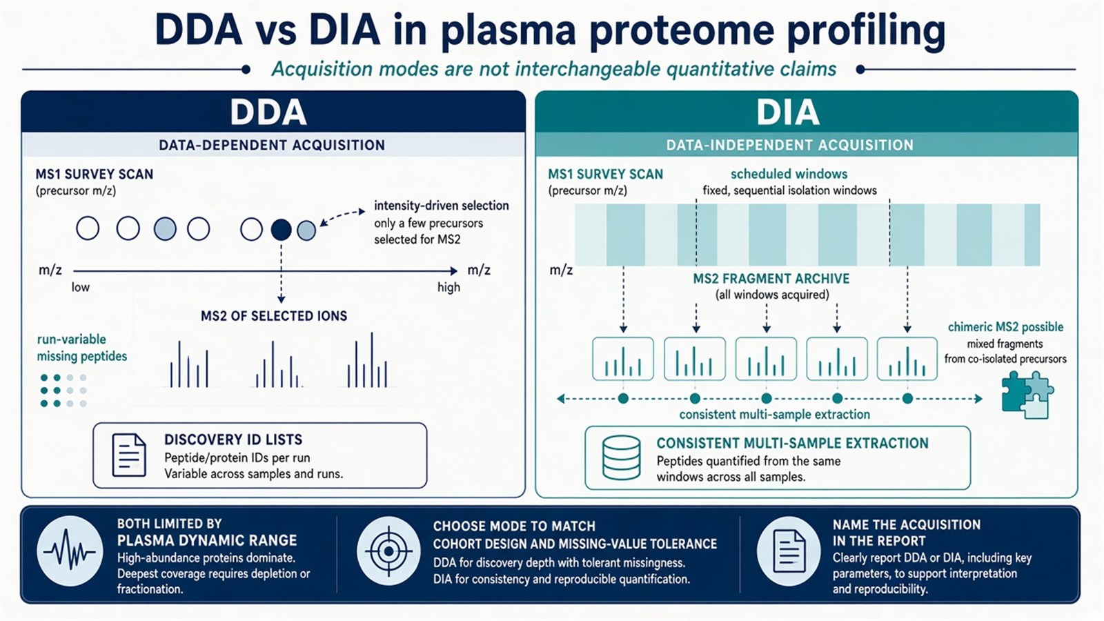
Figure 3. Plasma proteome profiling may acquire data as DDA with precursor selection or as DIA with scheduled windows; the two modes are not interchangeable names for one quantitative claim.
Bioinformatics After Profiling
Beyond acquisition, plasma proteome profiling requires computational analysis of the raw files. Database search or library extraction assigns peptides, protein inference summarizes those peptides, and quantitative matrices are then compared across the predefined groups. Differential expression analysis, functional enrichment, and pathway analysis are used to explore possible biological roles and disease associations. Enrichment reports overrepresentation relative to a defined background. It does not prove that a pathway is active in plasma or in a distant organ.
As cohort size grows, batch, collection site, and depletion kit lot become part of the experimental design. Bioinformatics refinement cannot repair unmatched anticoagulants or mixed serum and plasma labels.
Post-translational modification profiling is not included by adding a modification name to an undepleted DDA search. Phosphorylation, glycosylation, and other PTM studies in plasma need enrichment and a separate analytical plan.
Research Applications and Limits of Clinical Language
Plasma proteome profiling is widely used in research on pathological mechanisms, treatment monitoring, and candidate marker generation for cancer, cardiovascular, and metabolic models. Pre- versus post-treatment plasma in an oncology study can highlight proteins that change with exposure and may later be tested as pharmacodynamic or response candidates. Those applications remain hypothesis-generating until orthogonal assays and independent cohorts are in place. Variations in plasma proteins are not, by themselves, clinically validated biomarkers, and a profiling table does not inform a personalized treatment strategy without a separately qualified test.
Experimental Limits and Design Boundaries
Plasma proteome profiling does not equalize proteins from all organs, does not remove preanalytical variance, and does not convert missing values into biological absence. Immunoaffinity depletion improves access to some lower-abundance proteins and depletes others that were bound to the removed carriers. DDA missingness and DIA window interference remain acquisition-specific risks. Pathway maps drawn from a plasma differential list describe the circulating proteome, not tissue-localized mechanism.
Diagnostic cutoffs, companion-diagnostic claims, and guaranteed early detection sit outside ordinary research plasma proteome profiling. Once the matrix, high-abundance handling, and quantitative claim are defined, the project can be scoped. Researchers can review the MtoZ Biolabs Plasma Proteomics Analysis Service for project-specific sample evaluation, feasibility assessment, and workflow planning.
Frequently Asked Questions
1. Is plasma proteome profiling the same as serum proteomics?
No. Plasma is collected with an anticoagulant and retains coagulation-related proteins. Serum is collected after clotting, which changes the measurable protein set. The two matrices should not be mixed in one quantitative model.
2. Must high-abundance proteins always be depleted?
No. Depletion can improve detection of some lower-abundance proteins and can also remove bound partners. Undepleted DIA or other undepleted designs keep native abundance structure at the cost of dynamic-range coverage. The choice belongs in the methods record.
3. Do DDA and DIA both quantify the plasma proteome automatically?
They can support relative protein-level trends when identification, normalization, and QC are declared. They do not report calibrated plasma concentration without standards.
4. Can plasma profiling diagnose cancer or cardiovascular disease?
Not by itself. Discovery tables can prioritize candidates. Diagnostic use requires a separately validated assay and clinical study framework.
5. Does pathway enrichment of plasma proteins prove a tissue mechanism?
No. Enrichment summarizes overrepresentation in the circulating proteome relative to a background. It does not localize activity to an organ.
Reference
- Anderson N.L., Anderson N.G. (2002). The human plasma proteome: history, character, and diagnostic prospects. Mol. Cell. Proteomics, 1, 845-867. https://doi.org/10.1074/mcp.R200007-MCP200
- Geyer P.E., Holdt L.M., Teupser D., Mann M. (2017). Revisiting biomarker discovery by plasma proteomics. Mol. Syst. Biol., 13, 942. https://doi.org/10.15252/msb.20156297
- Gillet L.C., Navarro P., Tate S., et al. (2012). Targeted data extraction of the MS/MS spectra generated by data-independent acquisition: a new concept for consistent and accurate proteome analysis. Mol. Cell. Proteomics, 11, O111.016717. https://doi.org/10.1074/mcp.M111.016717
