



Application and analysis of TMT technology in proteomics means putting an isobaric tandem mass tag on a closed set of protein samples and reading relative change from reporter ions in one liquid chromatography-tandem mass spectrometry (LC-MS/MS) run (Thompson et al. 2003). The application is the proteomics experiment that can wear that label: a global multiplex contrast, a modification-enriched peptide set, an affinity eluate, or a tissue panel that is already in hand. The analysis is the reporter-ion protein table, with peptide support and the plex map attached. Pathway maps and biomarker stories are interpretations of that table. They are not a second TMT measurement. Search titles such as The Application of TMT Technology in Proteomics point to the same experiment class. They do not name a method that compares chemically different tag masses at MS1.
Researchers who already have defined samples, comparison groups, or a specific project objective can also review the MtoZ Biolabs TMT Quantitative Proteomics Analysis Service for project-specific feasibility and analysis planning.
What TMT Application and Analysis Can Claim
A TMT application starts from a comparison that can be frozen before labeling. After identification and reporter extraction, a higher protein value can support a relative statement about that protein across channels. Functional annotation and network figures can organize the list. They cannot repair incomplete labeling or co-isolation (Ting et al. 2011).
TMT tags are isobaric. They do not have different intact masses that separate channels in the survey scan. Sequence ions identify the peptide. Reporter ions quantify the channels.
Researchers who need to determine whether isobaric tagging matches a study can refer to TMT-Based Proteomics for more detailed guidance.
TMT application in proteomics is a closed multiplex experiment; TMT analysis is the reporter-ion table from that experiment.
Proteomics Experiments That Can Wear a TMT Label
Global multiplex relative discovery
Treatment, genotype, dose, or a short time course can use TMT when every sample enters one kit and peptides can be labeled at amines. This is the default proteomics application of the technology. It is not a large-cohort method merely because a kit holds 16 or 18 channels. Mixing still closes the block.
Modification-enriched peptides
A phosphopeptide or other enrichment can be labeled after digestion when amines remain available. The reporter then tracks the enriched peptide across channels. That is not the same number as protein abundance, and it is not site occupancy unless the unmodified counterpart and the experimental design support that claim. Signal-transduction language does not turn a phospho-TMT table into a pathway mechanism.
Affinity eluates and immunoprecipitates
Bait-versus-control or condition-versus-condition eluates can share one plex when yields are matched well enough to label. The application is relative change of proteins recovered with the bait. It is not a complete interactome, and leftover channels should not be filled with unrelated global digests.
Tissue or cell-type panels already in hand
Different tissues or cell types can be compared when the set is complete before labeling. Tissue-specific expression in a TMT table is still a relative ratio inside that panel. It is not a copy-number atlas across proteins, and it is not comparative genomics. A later tissue that arrives after mixing needs a new plex or an unlabeled run.
Long-term disease-progression series that will keep enrolling are a poor TMT application. Those designs need a bridge plan across plexes or unlabeled acquisition.
For case-control or drug-response studies that are being read as biomarker or target claims, researchers can refer to Application of TMT-Based Quantitative Proteomics in Biomarker Discovery for more detailed guidance.
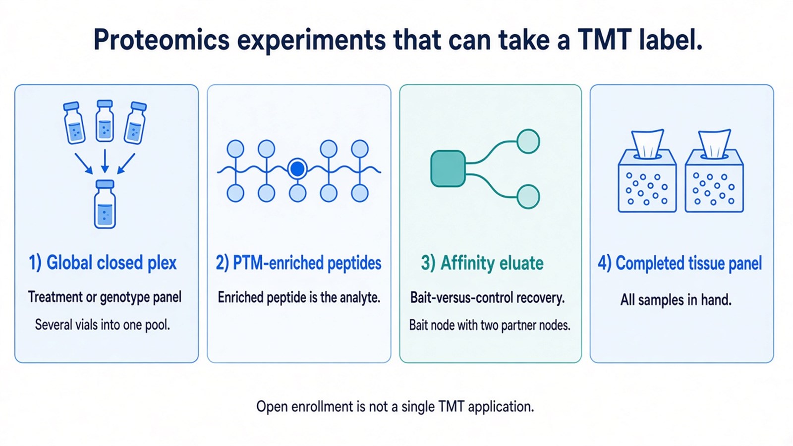
Figure 1. TMT technology applies to proteomics experiments that can be frozen into one kit; enrichment and affinity work still report relative channel change of the labeled analyte.
What TMT Analysis Includes and What It Adds Afterward
Analysis of a TMT proteomics experiment starts with a linked identification and reporter trail: the same search, the same feature map, and the stated impurity correction. Alignment in the unlabeled sense is not the TMT quantitative step. Channels already share one chromatogram. Peak identification here means assigning the peptide and extracting its reporters, not reconstructing independent LFQ runs.
A protein-level ratio can be summarized from unique peptides that carry the tag. Shared peptides, missed cleavage, and missing reporters belong in the report. Fold-change cutoffs and p-values are statistical choices. They are not built into TMT technology.
Bioinformatics steps that follow, including gene-ontology tables and pathway diagrams, interpret the differential list. They are useful when the list is already supported. They are not the analysis that makes TMT quantitative. Metabolic-regulation or drug-mechanism chapters written from those diagrams overstate the measurement.
In MtoZ isobaric-labeling offerings, TMT proteomics files are typically acquired in data-dependent acquisition (DDA) so reporter ions can be read. Service configuration is not an academic rule that other laboratories must copy.
Proteomics application |
What TMT analysis can support |
What the same files do not support |
|---|---|---|
Global closed contrast |
Relative protein change across channels |
An amount or a growing cohort |
PTM-enriched TMT |
Relative change of the enriched peptide |
Protein abundance or occupancy by default |
IP or affinity TMT |
Relative change in the recovered set |
A complete interaction network |
Frozen tissue panel |
Relative tissue-to-tissue ratios in that plex |
Comparative genomics or a copy-number map |
Pathway or network overlay |
Organization of a supported list |
Proof of mechanism or a biomarker |
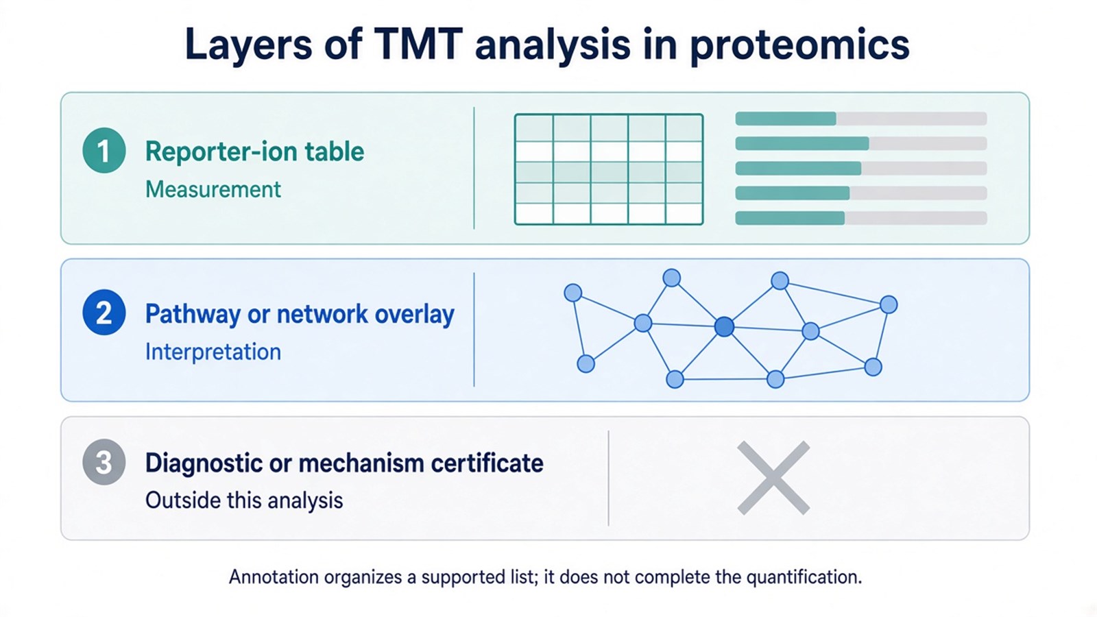
Figure 2. TMT analysis stops at a supported reporter-ion table; pathway figures interpret that table, and diagnostic or mechanism certificates require a different claim.
TMT analysis in proteomics reports relative channel ratios; it does not complete biology by annotation.
Frequently Asked Questions
1. Is application of TMT technology the same as TMT biomarker discovery?
Biomarker or drug-target language is one reading of a closed TMT contrast. The proteomics application is broader: any experiment that can be labeled, mixed, and compared as reporters. A differential protein is still a candidate, not a marker.
2. Does TMT analysis include pathway enrichment?
It can, as an optional interpretation after the ratio table exists. Enrichment is not required to call the experiment quantitative, and it does not validate labeling or isolation quality.
3. Can TMT monitor disease progression over a long clinical series?
Only if every time point is in hand, or if later points are run as new plexes with a stated bridge plan. Open enrollment is not a single TMT application.
4. Do TMT tags have different masses that the mass spectrometer uses to separate samples?
No. Tags in one kit are isobaric at MS1. Channels become visible when reporter ions are released on fragmentation.
Once the proteomics experiment type, closed sample set, and expected reporter-level readout are defined, the project can be evaluated as a TMT application. Researchers can review the MtoZ Biolabs TMT Quantitative Proteomics Analysis Service for project-specific sample evaluation, feasibility assessment, and workflow planning.
