


The specific steps for DSS protein crosslinking are two routes, not one general list. Route A treats a protein or a lysate that is already in an amine-free buffer. Route B treats intact cells, then opens them after the quench. Disuccinimidyl suberate is already an N-hydroxysuccinimide (NHS) ester. It is not activated at room temperature. Lysis is not step 1 when the proteins are still inside the cell. Dithiothreitol (DTT) does not hold proteins in a native state for crosslinking.
The six steps below are the protein-or-lysate sequence. A DMSO stock, immediate use, an amine quench, and one named assay stay in that sequence. An hours-long incubation, a spin that is said to remove leftover DSS, and a second mass-spectrometry heading that does not name the assay do not. What this chemistry can report is discussed in DSS Protein Crosslinking Experiment.
If a DSS route is already chosen and the readout is a residue-pair list, researchers can review the MtoZ Biolabs Crosslinking Mass Spectrometry Protein Interaction Analysis Service for project-specific feasibility and analysis planning.
Choose the DSS Route Before the First Step
DSS is membrane-permeable. Adding it to a culture or a washed cell suspension is a different first move from adding it to a purified complex. BS3 is the sulfonated analog that stays outside membranes; it does not replace DSS on Route B. A two-protein tube is Route A. What that tube must contain is explained in What Reagents Are Required for In Vitro Cross-Linking of Two Purified Proteins.
If the proteins are intracellular and the question is an in-cell link, do not extract them before the pulse. Lysis after an unquenched NHS ester continues the reaction in a new solvent. Whether lysis is the next move is discussed in Is Cell Lysis the Next Step After Protein Crosslinking? What Is the Best Lysis Buffer to Use.
A no-DSS arm is processed through the same buffer, quench, and assay. Changing only the treated sample mixes a handling effect with a reagent effect.
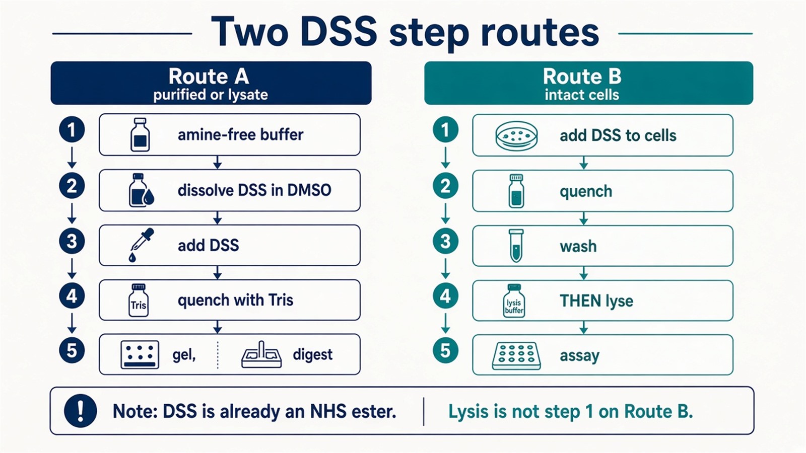
Figure 1. Route A starts from an amine-free protein or lysate. Route B adds DSS to intact cells and lyses after the quench. DSS is already an NHS ester.
Choose Route A or Route B before dissolving the solid. A procedure that starts with cell lysis is the wrong first step for in-cell DSS.
Route A: Six Steps for a Protein or Lysate
These steps follow this order.
1. Protein preparation
2. DSS preparation
Dissolve the solid in dry DMSO and dilute into the aqueous sample immediately. The ester hydrolyzes in water. Final DMSO is kept low so that the solvent spike is not the main denaturant. A molar excess relative to protein is one dosing style. A micromolar-to-millimolar titration is the other. Neither is a universal excess.
3. Crosslinking reaction
Add the dissolved DSS, mix, and incubate. About thirty minutes at room temperature is a common starting window. Ice-cold pulses are used when hydrolysis and over-linking need to be slowed. Leaving the ester in water for hours is not a more complete architecture. Extra time is mostly hydrolysis.
4. Reaction quenching
Add Tris or another amine in excess to consume leftover NHS ester. The quench stops further amine marking. It does not prevent unintended sites that already formed during the pulse. The order after the quench is described in How to Handle Samples After Adding Crosslinkers in Protein Crosslinking.
5. Cleanup, not a clarifying spin
Unreacted DSS and hydrolyzed NHS products are removed by buffer exchange, precipitation, or a later digest cleanup. Centrifugation pellets debris or cells. It does not subtract leftover crosslinker from a clear supernatant. A stained gel is optional. Many residue-pair datasets come from an in-solution digest.
6. One named assay
The last step is the readout, not a second copy of mass spectrometry. A gel shift, an immunoblot, or liquid chromatography–tandem mass spectrometry (LC-MS/MS) of linked peptides answer different questions. Searching the digest as ordinary peptides names proteins. It does not report DSS pairs.
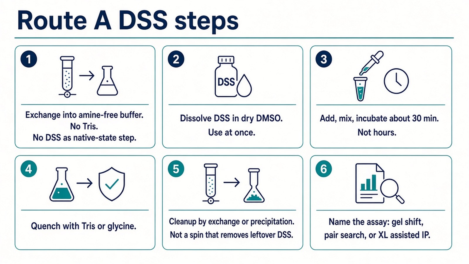
Figure 2. Route A order. DTT is not a native-state step. A spin does not remove leftover DSS. The last step names one assay.
Route B: Add DSS to Intact Cells, Then Lyse
What Is the Protocol for Crosslinking-Based Protein Interaction Analysis?.
What These DSS Steps Do Not Show
DSS does not become an NHS ester when the room is warm. The solid is already that ester. Room temperature is an incubation choice, not an activation. The steps do not establish a native-state conformation or a complete interface. A band that became larger is a mass shift. It is not a site.
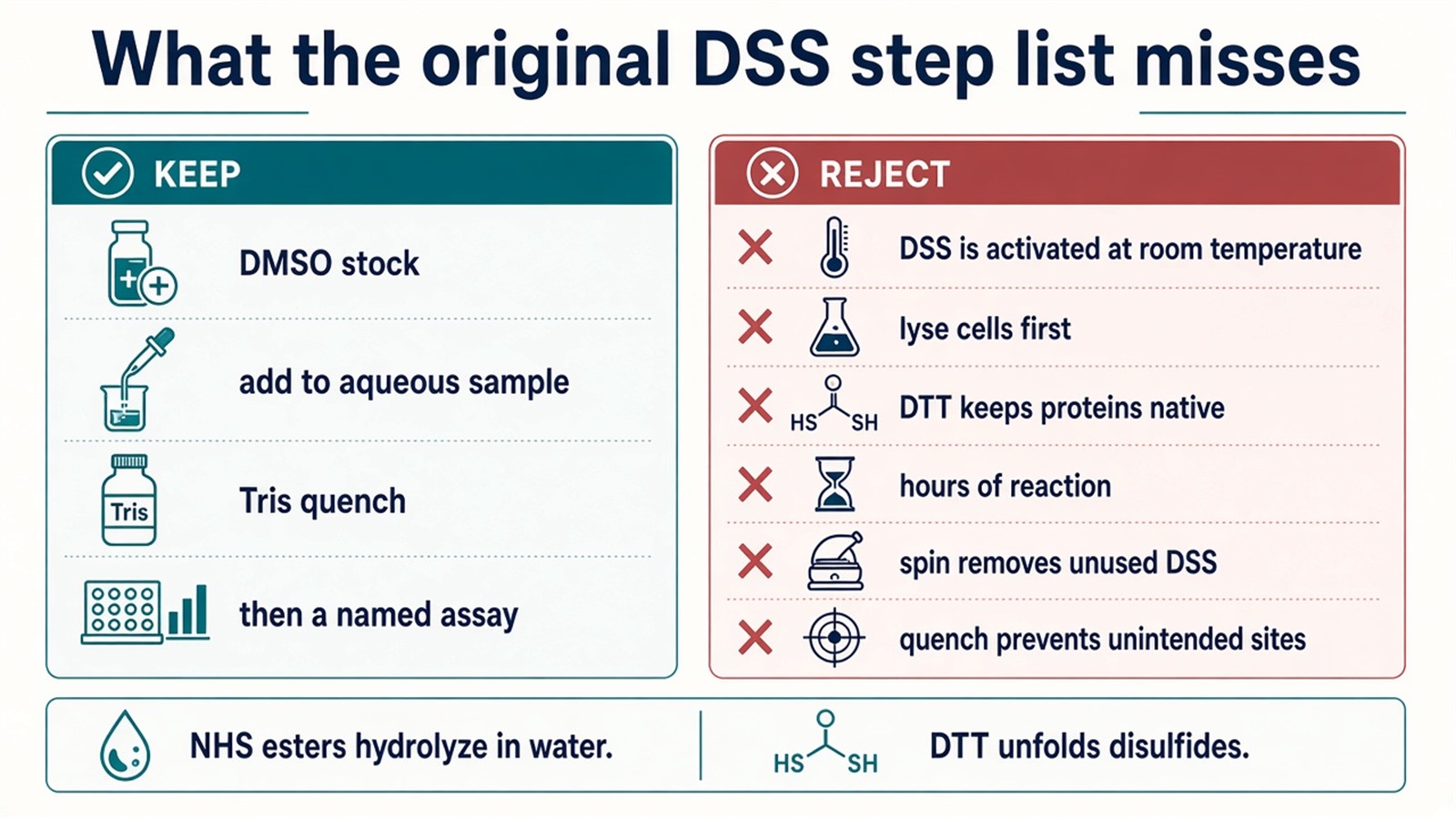
Figure 3. Keep: DMSO stock, aqueous pulse, amine quench, named assay. Not granted: activation at room temperature, lyse first, DTT as native, hours, spin cleanup.
What One DSS Procedure Cannot Establish
One DSS procedure covers one route, one dose, one quench, and one assay. It does not inventory every amine contact, and it does not set a diagnostic cutoff unless a separately validated assay is in place. Changing the route after the first failed gel does not convert Route A into Route B.
How to Plan a DSS Experiment
A usable DSS plan names the route, the amine-free or in-cell condition, the quench, and the assay that will read the pulse, before the first DMSO spike. When the readout is linked peptides, that plan is a crosslinking mass spectrometry study rather than a general fixation list. The interpretation still depends on the route, the dose, and the quench.
If the question is a cellular partner list rather than a DSS pair or a gel shift, do not force the sample through this step list. Researchers who need a partner-list study can review the MtoZ Biolabs Protein-Protein Interaction Analysis Service for project-specific sample evaluation, feasibility assessment, and workflow planning.
Frequently Asked Questions
1. What are the specific steps for DSS protein crosslinking?
Two routes. Route A: amine-free protein or lysate, DMSO stock, pulse, quench, cleanup, named assay. Route B: add DSS to intact cells, quench, then lyse.
2. Should intracellular proteins be lysed before DSS is added?
Not on the in-cell route. Lysis is after the quench.
3. Does DTT keep proteins native for DSS crosslinking?
No. Reducing a disulfide can unfold the domain that bond stabilizes. It is a perturbation, not a native-state step.
4. Does a Tris quench prevent unintended crosslinking sites?
No. It consumes leftover NHS ester. Sites that already formed remain.
5. Can centrifugation remove unreacted DSS?
No. A spin pellets debris. Leftover reagent is removed by exchange, precipitation, or later cleanup.
Reference
- Leitner A., Walzthoeni T., Kahraman A., et al. (2014). Probing native protein structures by chemical cross-linking, mass spectrometry, and bioinformatics. Nat. Protoc., 9, 120-135. https://doi.org/10.1038/nprot.2013.168
- Merkley E.D., Rysavy S., Kahraman A., et al. (2014). Distance restraints from crosslinking mass spectrometry: mining a molecular dynamics database to evaluate lysine-lysine distances. Protein Sci., 23, 747-759. https://doi.org/10.1002/pro.2458
- O'Reilly F.J., Rappsilber J. (2018). Cross-linking mass spectrometry: methods and applications in structural, molecular and systems biology. Nat. Struct. Mol. Biol., 25, 1000-1008. https://doi.org/10.1038/s41594-018-0147-0
- Yu C., Huang L. (2018). Cross-linking mass spectrometry: an emerging technology for interactomics and structural biology. Anal. Chem., 90, 144-165. https://doi.org/10.1021/acs.analchem.7b04431
- Sinz A. (2018). Cross-linking/mass spectrometry for studying protein structures and protein-protein interactions: where are we now and where should we go from here? Angew. Chem. Int. Ed., 57, 6390-6396. https://doi.org/10.1002/anie.201709559
