Western Blotting Quantitative Service
MtoZ Biolabs provides Western Blotting Quantitative Service for target protein detection and relative expression analysis using SDS-PAGE, membrane transfer, antibody recognition, and band-intensity quantification.
Chemiluminescence or fluorescence imaging can be selected according to assay conditions, with normalization and signal-range assessment supporting quantitative comparison across samples.
- Relative quantification of predefined target proteins.
- Band densitometry with normalization and quality control.
- Supports cells, tissues, biofluids, and purified proteins.
What Is Western Blot Quantification?
Western blot quantification compares the relative expression levels of proteins across different samples by analyzing the signal intensity of target protein bands detected with specific antibodies. Proteins are first separated by molecular weight using SDS-PAGE and transferred to a PVDF or nitrocellulose membrane. The target protein is then recognized by a specific primary antibody and a detection-labeled secondary antibody.
After the imaging system records chemiluminescent or fluorescent signals, band intensity can be measured by band densitometry and corrected using a loading control, total protein, or another suitable normalization approach. Reliable relative quantification requires the target signal to remain within the linear detection range, while sample loading, membrane transfer, antibody incubation, and imaging conditions should be kept as consistent as possible.
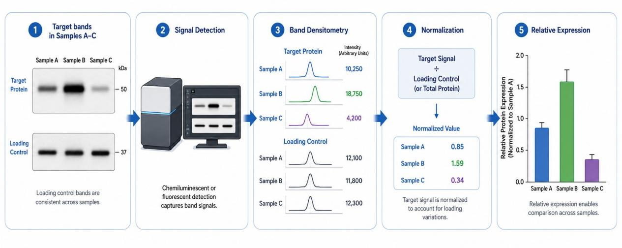
Figure 1. Western Blot Quantification Principle.
Service at MtoZ Biolabs
MtoZ Biolabs provides Western Blotting Quantitative Service, covering sample information and target protein evaluation, protein extraction, SDS-PAGE, membrane transfer, antibody incubation, chemiluminescence or fluorescence imaging, and band densitometry. Depending on the assay design, target protein signals can be normalized and compared across groups. Western blot images, relative quantification results, experimental information, and analysis reports can also be provided.
When This Service Is a Good Fit
- Defined target proteins and suitable antibodies are available, and relative expression levels need to be compared across different samples or treatment conditions.
- Target protein band position and signal intensity need to be evaluated together to compare expression changes across different samples.
- Candidate proteins identified through discovery proteomics require supplementary antibody-based validation.
- Specific protein forms, such as phosphorylated proteins, are of interest and suitable modification-specific antibodies and assay designs are available.
Analysis Workflow
1. Sample and Target Protein Evaluation
Confirm sample type, target protein, group design, biological replicates, available sample amount, and antibody information.
2. Protein Extraction and Concentration Measurement
Extract total protein according to sample type and measure protein concentration to arrange consistent loading amounts.
3. SDS-PAGE and Membrane Transfer
Separate proteins by SDS-PAGE and transfer the proteins to a PVDF or nitrocellulose membrane.
4. Blocking and Antibody Incubation
After membrane blocking, incubate the membrane with a target protein-specific primary antibody and the corresponding secondary antibody.
5. Signal Detection and Imaging
Perform chemiluminescence or fluorescence imaging according to the detection system and control exposure conditions to avoid signal saturation.
6. Band Densitometry and Normalization
Measure target-band signal intensity, normalize the signal using a suitable loading control or total-protein approach, and compare relative expression across groups.
Why Choose MtoZ Biolabs?
1. One-Stop Analysis
MtoZ Biolabs integrates sample evaluation and protein extraction with Western blot analysis, imaging, densitometry, and result organization.
2. Antibody and Detection Condition Evaluation
Western blot detection conditions are evaluated according to the target protein, available antibody information, and sample condition, followed by appropriate antibody incubation and signal detection arrangements.
3. Multiple Sample Types
Protein extraction methods can be selected according to the characteristics of microbial samples, animal or plant tissues, cells, and other sample types, followed by Western blot analysis.
4. Results and Data Delivery
Western blot images, band intensity data, normalized relative expression results, experimental information, and analysis reports can be provided.
Sample Submission Requirements
|
Sample Status |
Suggested Information |
|
Cell / Tissue Samples |
Sample source, species, group design, storage condition, available sample amount, target protein, and available antibody information. |
|
Serum / Plasma and Other Biofluids |
Sample type, collection and storage conditions, available volume, group design, target protein, and expected abundance information, if available. |
|
Protein Extracts |
Extraction method, buffer system, protein concentration/total amount, storage condition, target protein, and antibody information. |
|
Purified Proteins or Other Samples |
Protein source, purity/concentration information, buffer system, storage condition, and detection target. |
The actual sample amount required depends on sample type, target protein abundance, antibody performance, the number of targets to be measured, and the loading design. Providing complete sample and antibody information during consultation helps evaluate Western blot quantification conditions and sample submission arrangements.
Applications
1. Protein Expression Comparison
Compare relative target protein expression across different experimental groups, treatment conditions, or time points.
2. Signaling Pathway Research
Detect signaling pathway-related proteins and changes in protein expression to provide protein-level experimental data for studies of pathway responses.
3. Treatment Response Studies
Compare changes in target proteins before and after drug treatment, stimulation, or other experimental interventions.
4. Protein Modification Research
When suitable modification-specific antibodies are available, compare relative changes in phosphorylated proteins or other specific protein forms across different samples or experimental conditions.
5. Candidate Protein Validation
Perform supplementary Western blot validation of candidate proteins identified through proteomics or other screening approaches.
Deliverables
1. Sample Processing and Western Blot Experimental Information
2. Raw or Processed Western Blot Images
3. Target Protein Band Intensity Data
4. Normalized Relative Expression Results
5. Between-Group Comparison Results and Necessary Statistical Information, Where Applicable
6. Analysis Report
FAQ
Q1: Why should signal saturation be avoided in Western blot quantification?
When band signals exceed the linear detection range of the imaging system, signal intensity no longer maintains an approximately linear relationship with protein amount. Quantitative comparison therefore requires suitable exposure conditions, with target bands kept within a comparable signal range whenever possible.
Q2: Should Western blot quantification use a loading-control protein or total-protein normalization?
The normalization approach should be selected according to sample type, target protein, and experimental design. A loading-control protein should remain relatively stable under the comparison conditions, while total-protein normalization can be used to assess protein loading across the entire lane. Suitability should be evaluated according to the experimental conditions before selecting the normalization approach.
Q3: Can multiple target proteins with different molecular weights be detected on the same membrane?
The feasibility can be evaluated. Suitability for detection on the same membrane depends on target protein molecular weights, antibody host species, detection method, and band separation. When signal interference among multiple target proteins is substantial, the detection sequence or experimental arrangement may need to be adjusted.
Q4: Why cannot Western blot band intensity be used directly as an absolute protein concentration?
Band intensity in conventional Western blot analysis is affected by antibody-binding efficiency, membrane transfer, the detection system, and imaging conditions and is generally used for relative or semi-quantitative comparison across samples. Absolute concentration measurement requires additional standards, calibration curves, and a corresponding experimental design.
For Western blot quantitative analysis, sample type, species, group design, biological replicates, target protein, available antibody information, and available sample amount can be provided during consultation. The MtoZ Biolabs technical team will use the sample conditions and detection targets to confirm the experimental arrangement, sample submission requirements, and quantitative analysis approach.
