



Protein-protein interaction analysis asks which proteins remain with a defined partner after a stated test and matched controls. Far-Western blot answers that question on a membrane. Prey proteins are separated, transferred, and often renatured. A labeled bait is then used as the probe. A band means the probe stayed on that molecular-weight window after wash (Wu et al. 2007). Prefer this path when a purified bait exists and the claim is overlay retention on a size-resolved prey. Reassess when membrane renaturation is unnamed for a fold-dependent pair, probe-only or blocked-prey arms are missing, or the band is written as proven direct binding or a dissociation constant.
Researchers who already have a purified bait probe, a prey lysate or recombinant target, and a written overlay claim can also review the MtoZ Biolabs Far-Western Blot Analysis Service for project-specific feasibility.
Membrane Renaturation Before Any Band Is Kept
Far-Western blot is a membrane overlay assay, not an antibody Western and not a solution binding measurement. Prey proteins are usually resolved by SDS-PAGE, transferred to a membrane, and then offered a labeled bait as the probe. SDS-PAGE unfolds most proteins. If the intended contact needs fold, the membrane must be renatured before the probe is added. A skipped renaturation step can erase a real pair or leave a denatured surface that binds the probe in a new, non-native way.
Renaturation is a system choice, not a decoration. Name whether the blot was left denatured, partially refolded, or taken through a written renaturation series. Name whether the pair is expected to survive that path. A domain that binds as a linear motif can be readable on a denatured blot. A pair that needs a folded pocket is not. Do not write a denatured overlay as physiological binding. Do not write a refolded blot as proof that the cellular fold returned.
The system is readable when three things are named. First, a purified bait can be labeled without destroying the contact surface. Second, the prey can be seen at a stated load on the same membrane class. Third, the written claim is overlay retention, not an interactome and not a modification map. Far-Western does not report other proteins in a complex, and it does not report the modification form of the prey.
The system is a poor first choice when no purified bait exists, when the pair is known to need a fold that the blot cannot restore, when the prey is too scarce to appear after transfer, or when the next sentence is endogenous co-recovery. Capture after wash belongs on Co-IP or a pull-down, compared in What Are the Main Differences Between GST-Pull Down and Co-IP?.
Far-Western blot reports candidate overlay retention after a labeled bait meets a membrane-bound prey. It does not certify a binary protein-protein contact in solution.
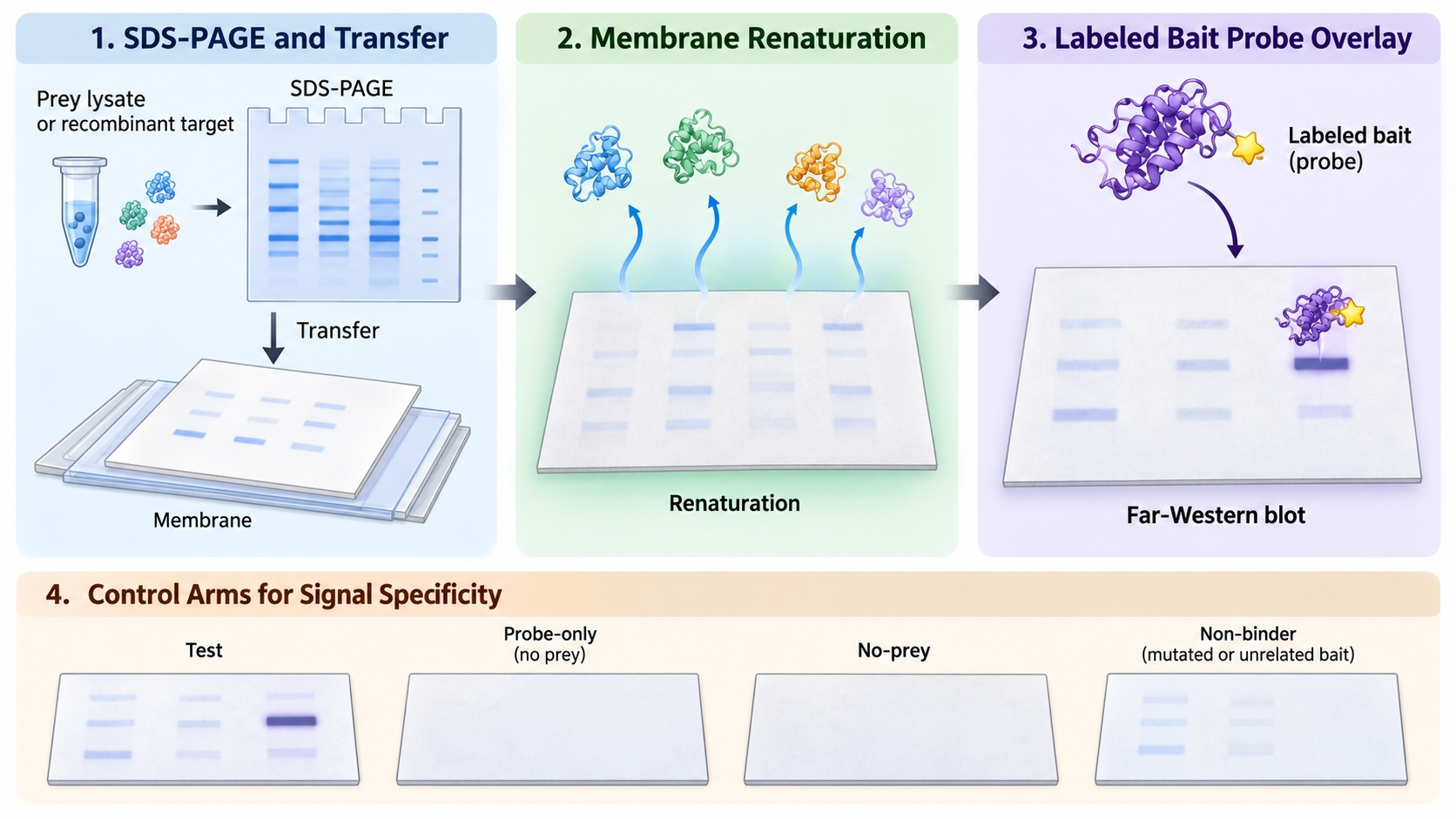
Figure 1. Far-Western blot workflow from SDS-PAGE and transfer through membrane renaturation to labeled bait probe overlay.
Probe Binding and Signal Specificity
Probe binding is the numerator of Far-Western blot. Signal specificity is the denominator. Neither is optional after one lane looks dark.
The probe is a labeled bait, not a primary antibody against the prey. Labeling must leave the contact surface usable. A biotin, epitope, or enzyme tag that sits on the interface can give a blank blot that looks like a negative result. Prey load must be visible by stain or a parallel Western against the prey. A missing prey band is not evidence that the pair failed.
Controls travel on the same membrane recipe. A probe-only or no-prey lane shows reagent that sticks to empty membrane. A blocked or mutated bait shows whether the mark still appears after the intended surface is broken. An unrelated bait that carries the same label shows chemistry that follows the tag. A no-renaturation arm, when fold is required, shows whether the signal depends on the refolding step. Unmatched probe concentration or unmatched exposure between test and control makes the contrast unreadable.
Intensity can compare the same prey across matched lanes on one blot. Intensity does not become a dissociation constant. Grayscale mixes prey amount, transfer, renaturation, probe activity, and exposure. Those variables do not rearrange into Kd.
|
System check |
What a pass looks like |
What the analysis cannot say |
|---|---|---|
|
Membrane renaturation |
Named denature or refold path matched to the pair |
Native cellular binding |
|
Probe binding |
Active labeled bait and a visible prey load |
A missing band as proof the pair is absent |
|
Signal specificity |
Probe-only, no-prey, and a non-binder on the same recipe |
Every dark lane is a partner |
|
Result boundary |
Candidate overlay retention under those arms |
Proven direct binding, a Kd, or gene regulation |
A practical path is:
- Write the overlay sentence the blot must support.
- Name SDS-PAGE or native separation, transfer, and the renaturation choice.
- Confirm prey load and probe activity before scoring darkness.
- Run probe-only and a non-binder with the same recipe.
- Send a passing pair to an orthogonal pull-down, Co-IP, or solution pair assay.
Signal specificity ranks a Far-Western band versus probe-only and non-binder arms. It does not certify a binary protein-protein contact or a Kd.
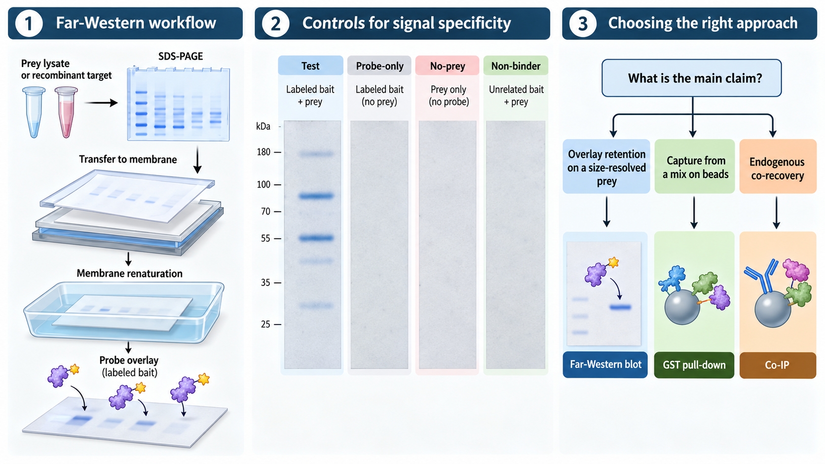
Figure 2. Control arms used to assess signal specificity in a Far-Western blot.
Validation Boundary and Common Biases
The common bias is writing Far-Western blot as a highly specific direct-binding proof because the probe is a protein rather than an antibody. Another bias is skipping renaturation because the protocol looks like an ordinary Western. A third is reading pixel intensity as affinity.
A band can be a retained pair, a denatured surface that newly sticks to the probe, or leftover label on a crowded molecular-weight window. Far-Western blot does not separate those classes by itself. It also does not show whether a third protein sat between bait and prey before SDS-PAGE. After denaturation, that third protein is gone. The overlay can still look binary.
Do not write a successful blot as proof of direct binding. Do not write the same blot as a Kd. Do not write a membrane or nuclear lysate as proof that the pair acts in that compartment. Do not write a dark band as gene regulation or a drug target. Prefer Far-Western blot when a purified bait exists and several lysates or mutants must be scored on one membrane. Prefer a GST pull-down when the next sentence is capture from a mix on beads, reviewed under GST Pull Down Assay Service. Prefer Co-IP or IP-MS when the next sentence is endogenous co-recovery, reviewed under Protein-Protein Interaction Analysis.
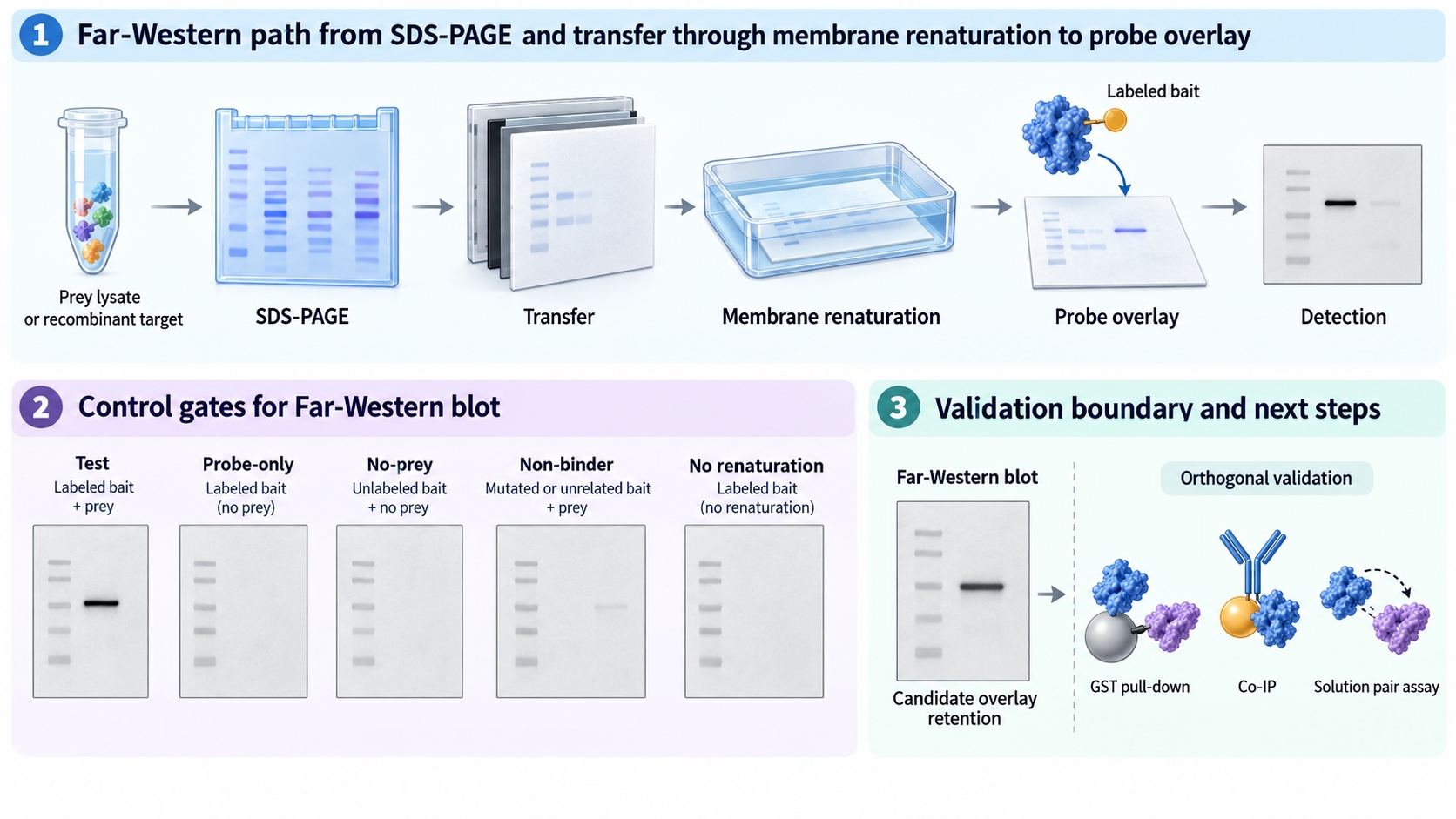
Figure 3. Far-Western blot identifies candidate overlay retention that can be followed by orthogonal validation.
Once the bait probe, prey load, renaturation path, and negative arms are defined, the project can be scoped as a Far-Western overlay test. Researchers can review the MtoZ Biolabs Far-Western Blot Analysis Service for sample evaluation and control planning.
Frequently Asked Questions
1. Does a Far-Western band prove two proteins bind directly?
No. The band reports that a labeled bait stayed on a membrane-bound prey after wash. Direct contact in solution needs an orthogonal pair assay.
2. Do I need membrane renaturation?
Name it. If the pair needs fold, skip that step and the blot can go blank or create a non-native band. If the contact is a linear motif, a denatured blot may still be readable.
3. Can band intensity be read as Kd?
No. Intensity mixes load, transfer, renaturation, probe activity, and exposure. It can compare the same prey across matched lanes. It does not return a dissociation constant.
4. Is Far-Western blot the same as a Western blot?
No. A Western uses an antibody to find a named prey. Far-Western uses a labeled bait as the probe. Antibody performance on the prey does not replace probe-only controls.
5. When should this assay move to pull-down or Co-IP?
Move to a pull-down when the next written sentence is bead capture from a mix. Move to Co-IP when the next sentence is endogenous co-recovery. Keep the Far-Western shortlist as the input, not as the proof.
Reference
- Y. Wu, Q. Li, X.Z. Chen (2007). Detecting protein-protein interactions by Far western blotting. Nat. Protoc., 2, 3278-3284. https://doi.org/10.1038/nprot.2007.459
